 Movie
Movie Controller

[English] 日本語
 Yorodumi
- PDB-1v84: Crystal structure of human GlcAT-P in complex with N-acetyllactos... -
+ Open data

- Basic information
| Entry | Database: PDB / ID: 1v84 | |||||||||
|---|---|---|---|---|---|---|---|---|---|---|
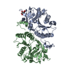
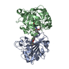
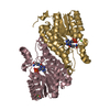
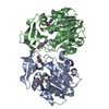
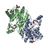
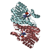
| Title | Crystal structure of human GlcAT-P in complex with N-acetyllactosamine, Udp, and Mn2+ | |||||||||
 Components Components | Galactosylgalactosylxylosylprotein 3-beta-glucuronosyltransferase 1 | |||||||||
 Keywords Keywords | TRANSFERASE / glycoprotein / glycocyltransferase / HNK-1 epitope | |||||||||
| Function / homology |  Function and homology information Function and homology informationgalactosylgalactosylxylosylprotein 3-beta-glucuronosyltransferase / galactosylgalactosylxylosylprotein 3-beta-glucuronosyltransferase activity / Glycosaminoglycan-protein linkage region biosynthesis / glycosaminoglycan biosynthetic process / chondroitin sulfate proteoglycan biosynthetic process / carbohydrate metabolic process / Golgi membrane / endoplasmic reticulum membrane / extracellular region / membrane / metal ion binding Similarity search - Function | |||||||||
| Biological species |  Homo sapiens (human) Homo sapiens (human) | |||||||||
| Method |  X-RAY DIFFRACTION / SYNCHROTRON / MOLECULAR REPLACEMENT / Resolution: 1.82 Å X-RAY DIFFRACTION / SYNCHROTRON / MOLECULAR REPLACEMENT / Resolution: 1.82 Å | |||||||||
 Authors Authors | Kakuda, S. / Shiba, T. / Ishiguro, M. / Tagawa, H. / Oka, S. / Kajihara, Y. / Kawasaki, T. / Wakatsuki, S. / Kato, R. | |||||||||
 Citation Citation | Journal: J.Biol.Chem. / Year: 2004 Title: Structural Basis for Acceptor Substrate Recognition of a Human Glucuronyltransferase, GlcAT-P, an Enzyme Critical in the Biosynthesis of the Carbohydrate Epitope HNK-1 Authors: Kakuda, S. / Shiba, T. / Ishiguro, M. / Tagawa, H. / Oka, S. / Kajihara, Y. / Kawasaki, T. / Wakatsuki, S. / Kato, R. | |||||||||
| History |
|
- Structure visualization
| Structure viewer | Molecule: MolmilJmol/JSmol |
|---|
- Downloads & links
-Download
| PDBx/mmCIF format | 1v84.cif.gz | 124.1 KB | Display | PDBx/mmCIF format |
|---|---|---|---|---|
| PDB format | pdb1v84.ent.gz | 93 KB | Display | PDB format |
| PDBx/mmJSON format | 1v84.json.gz | Tree view | PDBx/mmJSON format | |
| Others |  Other downloads Other downloads |
-Validation report
| Arichive directory | https://data.pdbj.org/pub/pdb/validation_reports/v8/1v84ftp://data.pdbj.org/pub/pdb/validation_reports/v8/1v84 | HTTPS FTP |
|---|
-Related structure data
| Related structure data |  1v82SC 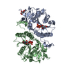 1v83C S: Starting model for refinement C: citing same article ( |
|---|---|
| Similar structure data |


-Links
 PDBj
PDBj


- Assembly
| Deposited unit | 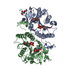
| ||||||||
|---|---|---|---|---|---|---|---|---|---|
| 1 |
| ||||||||
| Unit cell |
|
-Components
-Protein , 1 types, 2 molecules AB
| #1: Protein | Mass: 29038.408 Da / Num. of mol.: 2 / Fragment: catalytic domain Source method: isolated from a genetically manipulated source Source: (gene. exp.) Homo sapiens (human) / Plasmid: pET-28a(+) / Production host:  References: UniProt: Q9P2W7, galactosylgalactosylxylosylprotein 3-beta-glucuronosyltransferase |
|---|
-Sugars , 2 types, 2 molecules
| #2: Polysaccharide | beta-D-galactopyranose-(1-4)-2-acetamido-2-deoxy-alpha-D-glucopyranose / N-acetyl-alpha-lactosamine  Source method: isolated from a genetically manipulated source Details: oligosaccharide / References: N-acetyl-alpha-lactosamine |
|---|---|
| #3: Polysaccharide | beta-D-galactopyranose-(1-4)-2-acetamido-2-deoxy-beta-D-glucopyranose Source method: isolated from a genetically manipulated source |
-Non-polymers , 4 types, 365 molecules 


| #4: Chemical |  Mass: 54.938 Da / Num. of mol.: 2 / Source method: obtained synthetically / Formula: Mn Mass: 54.938 Da / Num. of mol.: 2 / Source method: obtained synthetically / Formula: Mn#5: Chemical | ChemComp-TLA / |  Mass: 150.087 Da / Num. of mol.: 1 / Source method: obtained synthetically / Formula: C4H6O6 Mass: 150.087 Da / Num. of mol.: 1 / Source method: obtained synthetically / Formula: C4H6O6#6: Chemical |  Type: RNA linking / Mass: 404.161 Da / Num. of mol.: 2 / Source method: obtained synthetically / Formula: C9H14N2O12P2 / Comment: UDP*YM Type: RNA linking / Mass: 404.161 Da / Num. of mol.: 2 / Source method: obtained synthetically / Formula: C9H14N2O12P2 / Comment: UDP*YM#7: Water | ChemComp-HOH / | Mass: 18.015 Da / Num. of mol.: 360 / Source method: isolated from a natural source / Formula: H2O |
|---|
-Experimental details
-Experiment
| Experiment | Method: X-RAY DIFFRACTION / Number of used crystals: 1 |
|---|
- Sample preparation
| Crystal | Density Matthews: 2.61 Å3/Da / Density % sol: 52.5 % |
|---|---|
| Crystal grow | Temperature: 289 K / Method: vapor diffusion, hanging drop Details: PEG5000MME, di-sodium tartrate, VAPOR DIFFUSION, HANGING DROP, temperature 289K |
-Data collection
| Diffraction | Mean temperature: 100 K |
|---|---|
| Diffraction source | Source: SYNCHROTRON / Site: Photon Factory  / Beamline: AR-NW12A / Wavelength: 0.974 Å / Beamline: AR-NW12A / Wavelength: 0.974 Å |
| Detector | Type: ADSC QUANTUM 210 / Detector: CCD / Date: Jun 12, 2003 |
| Radiation | Monochromator: Si(111) / Protocol: SINGLE WAVELENGTH / Monochromatic (M) / Laue (L): M / Scattering type: x-ray |
| Radiation wavelength | Wavelength: 0.974 Å / Relative weight: 1 |
| Reflection | Resolution: 1.82→50 Å / Num. all: 57144 / Num. obs: 57093 / % possible obs: 97.1 % / Rmerge(I) obs: 0.067 / Net I/σ(I): 10.4 |
| Reflection shell | Resolution: 1.82→1.89 Å / Rmerge(I) obs: 0.379 / Mean I/σ(I) obs: 2.8 / % possible all: 88.4 |
- Processing
| Software |
| |||||||||||||||||||||||||
|---|---|---|---|---|---|---|---|---|---|---|---|---|---|---|---|---|---|---|---|---|---|---|---|---|---|---|
| Refinement | Method to determine structure: MOLECULAR REPLACEMENT Starting model: 1V82 Resolution: 1.82→40 Å / Cross valid method: THROUGHOUT / σ(F): 0 / Stereochemistry target values: Engh & Huber
| |||||||||||||||||||||||||
| Refinement step | Cycle: LAST / Resolution: 1.82→40 Å
| |||||||||||||||||||||||||
| Refine LS restraints |
|
