 Movie
Movie Controller

[English] 日本語
 Yorodumi
- PDB-1v7h: Crystal Structures of Collagen Model Peptides with Pro-Hyp-Gly Se... -
+ Open data

- Basic information
| Entry | Database: PDB / ID: 1v7h | ||||||
|---|---|---|---|---|---|---|---|
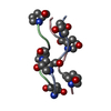
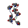
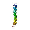
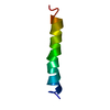
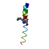
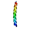
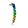
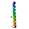
| Title | Crystal Structures of Collagen Model Peptides with Pro-Hyp-Gly Sequence at 1.26 A | ||||||
 Components Components | (Collagen like peptide) x 3 | ||||||
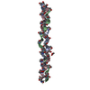
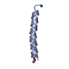
 Keywords Keywords | STRUCTURAL PROTEIN / Collagen / Triple-helix / Model peptide | ||||||
| Method |  X-RAY DIFFRACTION / SYNCHROTRON / MOLECULAR REPLACEMENT / Resolution: 1.25 Å X-RAY DIFFRACTION / SYNCHROTRON / MOLECULAR REPLACEMENT / Resolution: 1.25 Å | ||||||
 Authors Authors | Okuyama, K. / Hongo, C. / Fukushima, R. / Wu, G. / Narita, H. / Noguchi, K. / Tanaka, Y. / Nishino, N. | ||||||
 Citation Citation | Journal: Biopolymers / Year: 2004 Title: Crystal structures of collagen model peptides with Pro-Hyp-Gly repeating sequence at 1.26 A resolution: implications for proline ring puckering Authors: Okuyama, K. / Hongo, C. / Fukushima, R. / Wu, G. / Narita, H. / Noguchi, K. / Tanaka, Y. / Nishino, N. | ||||||
| History |
|
- Structure visualization
| Structure viewer | Molecule:  MolmilJmol/JSmol MolmilJmol/JSmol |
|---|
- Downloads & links
-Download
| PDBx/mmCIF format | 1v7h.cif.gz | 15.5 KB | Display | PDBx/mmCIF format |
|---|---|---|---|---|
| PDB format | pdb1v7h.ent.gz | 11 KB | Display | PDB format |
| PDBx/mmJSON format | 1v7h.json.gz | Tree view | PDBx/mmJSON format | |
| Others |  Other downloads Other downloads |
-Validation report
| Summary document | 1v7h_validation.pdf.gz | 354.6 KB | Display | wwPDB validaton report |
|---|---|---|---|---|
| Full document | 1v7h_full_validation.pdf.gz | 354.6 KB | Display | |
| Data in XML | 1v7h_validation.xml.gz | 1.8 KB | Display | |
| Data in CIF | 1v7h_validation.cif.gz | 2.3 KB | Display | |
| Arichive directory | https://data.pdbj.org/pub/pdb/validation_reports/v7/1v7hftp://data.pdbj.org/pub/pdb/validation_reports/v7/1v7h | HTTPS FTP |
-Related structure data

-Links
 PDBj
PDBj
- Assembly
| Deposited unit | 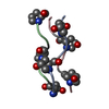
| ||||||||
|---|---|---|---|---|---|---|---|---|---|
| 1 | 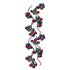
| ||||||||
| Unit cell |
| ||||||||
| Details | THE ENTIRE 30 RESIDUE LONG PEPTIDE CAN BE GENERATED FROM THE SUBMITTED ASYMMETRIC UNIT BY APPLYING THE FOLLOWING TRANSLATIONS (USING FRACTIONAL COORDINATES): CHAIN A: TRANSLATE RESIDUES 2 - 7 BY (001), AND RESIDUES 1-7 BY (002),(003),(004), AND RESIDUES 1-4 BY (005) CHAIN B: TRANSLATE RESIDUES 3 - 7 BY (001), AND RESIDUES 1-7 BY (002),(003),(004) AND RESIDUE 1-2 BY (005). CHAIN C: TRANSLATE RESIDUES 1 - 7 BY (001),(002),(003),(004), AND RESIDUES 1-3 BY (005). THIS WILL RESULT IN A MOLECULE WITH A TOTAL OF 90 RESIDUES, 30 IN EACH CHAIN. |
-Components
| #1: Protein/peptide | Mass: 609.630 Da / Num. of mol.: 1 / Source method: obtained synthetically Details: Pro-Hyp-Gly triplet is very popular in collagen sequence |
|---|---|
| #2: Protein/peptide | Mass: 665.693 Da / Num. of mol.: 1 / Source method: obtained synthetically Details: Pro-Hyp-Gly triplet is very popular in collagen sequence |
| #3: Protein/peptide | Mass: 649.693 Da / Num. of mol.: 1 / Source method: obtained synthetically Details: Pro-Hyp-Gly triplet is very popular in collagen sequence |
| #4: Water | ChemComp-HOH /  Mass: 18.015 Da / Num. of mol.: 49 / Source method: isolated from a natural source / Formula: H2O Mass: 18.015 Da / Num. of mol.: 49 / Source method: isolated from a natural source / Formula: H2O |
-Experimental details
-Experiment
| Experiment | Method: X-RAY DIFFRACTION / Number of used crystals: 1 |
|---|
- Sample preparation
| Crystal | Density Matthews: 1.81 Å3/Da / Density % sol: 31.96 % |
|---|---|
| Crystal grow | Temperature: 283 K / Method: vapor diffusion, hanging drop Details: PEG200, ACETIC ACID, VAPOR DIFFUSION, HANGING DROP, temperature 283K |
-Data collection
| Diffraction | Mean temperature: 100 K |
|---|---|
| Diffraction source | Source: SYNCHROTRON / Site: SPring-8  / Beamline: BL44XU / Wavelength: 0.9 Å / Beamline: BL44XU / Wavelength: 0.9 Å |
| Detector | Type: OXFORD PX210 / Detector: CCD / Date: Jun 11, 2002 / Details: MIRROR-MONOCHROMATOR |
| Radiation | Protocol: SINGLE WAVELENGTH / Monochromatic (M) / Laue (L): M / Scattering type: x-ray |
| Radiation wavelength | Wavelength: 0.9 Å / Relative weight: 1 |
| Reflection | Resolution: 1.24→20 Å / Num. obs: 3257 / Observed criterion σ(F): 1 / Rmerge(I) obs: 0.042 |
| Reflection shell | Resolution: 1.24→1.28 Å / Rmerge(I) obs: 0.057 / Mean I/σ(I) obs: 8.2 / Num. unique all: 290 / % possible all: 74 |
- Processing
| Software |
| |||||||||||||||||||||||||||||||||
|---|---|---|---|---|---|---|---|---|---|---|---|---|---|---|---|---|---|---|---|---|---|---|---|---|---|---|---|---|---|---|---|---|---|---|
| Refinement | Method to determine structure: MOLECULAR REPLACEMENT Starting model: (PRO-HYP-GLY)10 structure reported in V.NAGARAJAN, S.KAMITORI, K.OKUYAMA, J.BIOCHEM.,125, 310 (1999). Resolution: 1.25→10 Å / Num. parameters: 1396 / Num. restraintsaints: 1695 / Cross valid method: THROUGHOUT / σ(F): 4 / Stereochemistry target values: Engh & Huber
| |||||||||||||||||||||||||||||||||
| Refine analyze | Num. disordered residues: 0 / Occupancy sum hydrogen: 112 / Occupancy sum non hydrogen: 182 | |||||||||||||||||||||||||||||||||
| Refinement step | Cycle: LAST / Resolution: 1.25→10 Å
| |||||||||||||||||||||||||||||||||
| Refine LS restraints |
| |||||||||||||||||||||||||||||||||
| LS refinement shell | Resolution: 1.25→1.3 Å /
|
