[日本語] English
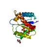
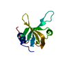
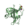
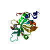
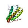
- PDB-1v5k: Solution structure of the CH domain from mouse EB-1 -

+
データを開く


IDまたはキーワード:

読み込み中...

-
基本情報

登録情報
データベース: PDB / ID: 1v5k
タイトルSolution structure of the CH domain from mouse EB-1
要素microtubule-associated protein, RP/EB family, member 1
キーワードSTRUCTURAL PROTEIN / PROTEIN BINDING / calponin homology (CH) domain / microtubule binding / adenomatosis polyposis coli binding protein / structural genomics / RIKEN Structural Genomics/Proteomics Initiative / RSGI
機能・相同性
機能・相同性情報


protein localization to astral microtubule / protein localization to mitotic spindle / cortical microtubule cytoskeleton / mitotic spindle astral microtubule end / Amplification of signal from unattached kinetochores via a MAD2 inhibitory signal / Mitotic Prometaphase / EML4 and NUDC in mitotic spindle formation / Resolution of Sister Chromatid Cohesion / The role of GTSE1 in G2/M progression after G2 checkpoint / protein localization to microtubule ...protein localization to astral microtubule / protein localization to mitotic spindle / cortical microtubule cytoskeleton / mitotic spindle astral microtubule end / Amplification of signal from unattached kinetochores via a MAD2 inhibitory signal / Mitotic Prometaphase / EML4 and NUDC in mitotic spindle formation / Resolution of Sister Chromatid Cohesion / The role of GTSE1 in G2/M progression after G2 checkpoint / protein localization to microtubule / RHO GTPases Activate Formins / Separation of Sister Chromatids / Loss of Nlp from mitotic centrosomes / Recruitment of mitotic centrosome proteins and complexes / Loss of proteins required for interphase microtubule organization from the centrosome / Recruitment of NuMA to mitotic centrosomes / Anchoring of the basal body to the plasma membrane / AURKA Activation by TPX2 / microtubule plus-end / mitotic spindle microtubule / Regulation of PLK1 Activity at G2/M Transition / cell projection membrane / attachment of mitotic spindle microtubules to kinetochore / microtubule bundle formation / microtubule plus-end binding / non-motile cilium assembly / protein localization to centrosome / negative regulation of microtubule polymerization / mitotic spindle pole / microtubule polymerization / establishment of mitotic spindle orientation / positive regulation of microtubule polymerization / protein serine/threonine kinase binding / cell projection / cell migration / microtubule cytoskeleton / microtubule / ciliary basal body / cell division / focal adhesion / centrosome / Golgi apparatus / identical protein binding
類似検索 - 分子機能
EB1, C-terminal / Microtubule-associated protein RP/EB / EB1, C-terminal domain superfamily / EB1-C terminal (EB1-C) domain profile. / EB1-like C-terminal motif / Calponin-like domain / Actin-binding Protein, T-fimbrin; domain 1 / Calponin homology (CH) domain / Calponin homology domain / CH domain superfamily ...EB1, C-terminal / Microtubule-associated protein RP/EB / EB1, C-terminal domain superfamily / EB1-C terminal (EB1-C) domain profile. / EB1-like C-terminal motif / Calponin-like domain / Actin-binding Protein, T-fimbrin; domain 1 / Calponin homology (CH) domain / Calponin homology domain / CH domain superfamily / Calponin homology (CH) domain profile. / Orthogonal Bundle / Mainly Alpha
類似検索 - ドメイン・相同性
: / Microtubule-associated protein RP/EB family member 1
類似検索 - 構成要素
生物種Mus musculus (ハツカネズミ)
手法溶液NMR / torsion angle dynamics, restrained molecular dynamics
データ登録者Tomizawa, T. / Kigawa, T. / Koshiba, S. / Inoue, M. / Yokoyama, S. / RIKEN Structural Genomics/Proteomics Initiative (RSGI)
引用ジャーナル: To be Published
タイトル: Solution structure of the CH domain from mouse EB-1
著者: Tomizawa, T. / Kigawa, T. / Koshiba, S. / Inoue, M. / Yokoyama, S.
履歴
登録2003年11月25日登録サイト: PDBJ / 処理サイト: PDBJ
改定 1.02004年5月25日Provider: repository / タイプ: Initial release
改定 1.12008年4月27日Group: Version format compliance
改定 1.22011年7月13日Group: Version format compliance
改定 1.32021年11月10日Group: Data collection / Database references / Derived calculations
カテゴリ: database_2 / pdbx_nmr_software ...database_2 / pdbx_nmr_software / pdbx_nmr_spectrometer / pdbx_struct_assembly / pdbx_struct_oper_list / struct_ref_seq_dif
Item: _database_2.pdbx_DOI / _database_2.pdbx_database_accession ..._database_2.pdbx_DOI / _database_2.pdbx_database_accession / _pdbx_nmr_software.name / _pdbx_nmr_spectrometer.model / _struct_ref_seq_dif.details
改定 1.42023年12月27日Group: Data collection / カテゴリ: chem_comp_atom / chem_comp_bond

-
構造の表示

構造ビューア分子:
MolmilJmol/JSmol

ダウンロードとリンク

-
集合体

登録構造単位
A: microtubule-associated protein, RP/EB family, member 1


分子量 (理論値)分子数
合計 (水以外)13,1191
ポリマ-13,1191
非ポリマー00
00
1


  • 登録構造と同一
  • 登録者が定義した集合体
タイプ名称対称操作
identity operation1_5551
NMR アンサンブル
データ基準
コンフォーマー数 (登録 / 計算)20 / 100structures with the least restraint violations, structures with the lowest energy, target function
代表モデルモデル #1lowest energy

-
要素

#1: タンパク質 microtubule-associated protein, RP/EB family, member 1 / adenomatosis polyposis coli binding protein Eb1


分子量: 13119.191 Da / 分子数: 1 / 断片: CH domain / 変異: L8Q/S9R / 由来タイプ: 組換発現 / 由来: (組換発現) Mus musculus (ハツカネズミ) / 解説: Cell-free protein synthesis / 遺伝子: RIKEN cDNA 2900038A16 / プラスミド: P021216-20 / 参照: GenBank: 7106301, UniProt: Q61166*PLUS

-
実験情報

-
実験

実験手法: 溶液NMR
NMR実験
Conditions-IDExperiment-IDSolution-IDタイプ
1113D 13C-separated NOESY
1213D 15N-separated NOESY

-
試料調製

詳細内容: 1.03mM CH domain U-15N, 13C; 20mM sodium phosphate; 100mM NaCl; 1mM d-DTT; 0.02% NaN3; 10% D2O
溶媒系: 90% H2O/10% D2O
試料状態イオン強度: 120mM / pH: 6 / : ambient / 温度: 298 K

-
NMR測定

放射プロトコル: SINGLE WAVELENGTH / 単色(M)・ラウエ(L): M
放射波長相対比: 1
NMRスペクトロメータータイプ: Bruker AVANCE / 製造業者: Bruker / モデル: AVANCE / 磁場強度: 800 MHz

-
解析

NMR software
名称バージョン開発者分類
XwinNMR2.6Brukercollection
NMRPipe20020425Delaglio, F.解析
NMRView5.0.4Johnson, B.A.データ解析
KUJIRA0.863Kobayashi, N.データ解析
CYANA1.0.7Guentert, P.構造決定
CYANA1.0.7Guentert, P.精密化
精密化手法: torsion angle dynamics, restrained molecular dynamics
ソフトェア番号: 1
代表構造選択基準: lowest energy
NMRアンサンブル
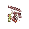
コンフォーマー選択の基準: structures with the least restraint violations, structures with the lowest energy, target function
計算したコンフォーマーの数: 100 / 登録したコンフォーマーの数: 20

+
万見について

-
お知らせ

-
2022年2月9日: EMDBエントリの付随情報ファイルのフォーマットが新しくなりました

EMDBエントリの付随情報ファイルのフォーマットが新しくなりました

  • EMDBのヘッダファイルのバージョン3が、公式のフォーマットとなりました。
  • これまでは公式だったバージョン1.9は、アーカイブから削除されます。

関連情報:EMDBヘッダ

外部リンク:wwPDBはEMDBデータモデルのバージョン3へ移行します

-
2020年8月12日: 新型コロナ情報

新型コロナ情報

URL: https://pdbj.org/emnavi/covid19.php

新ページ: EM Navigatorに新型コロナウイルスの特設ページを開設しました。

関連情報:Covid-19情報 / 2020年3月5日: 新型コロナウイルスの構造データ

+
2020年3月5日: 新型コロナウイルスの構造データ

新型コロナウイルスの構造データ

関連情報:万見生物種 / 2020年8月12日: 新型コロナ情報

外部リンク:COVID-19特集ページ - PDBj / 今月の分子2020年2月:コロナウイルスプロテーアーゼ

+
2019年1月31日: EMDBのIDの桁数の変更

EMDBのIDの桁数の変更

  • EMDBエントリに付与されているアクセスコード(EMDB-ID)は4桁の数字(例、EMD-1234)でしたが、間もなく枯渇します。これまでの4桁のID番号は4桁のまま変更されませんが、4桁の数字を使い切った後に発行されるIDは5桁以上の数字(例、EMD-12345)になります。5桁のIDは2019年の春頃から発行される見通しです。
  • EM Navigator/万見では、接頭語「EMD-」は省略されています。

関連情報:Q: 「EMD」とは何ですか? / 万見/EM NavigatorにおけるID/アクセスコードの表記

外部リンク:EMDB Accession Codes are Changing Soon! / PDBjへお問い合わせ

+
2017年7月12日: PDB大規模アップデート

PDB大規模アップデート

  • 新バージョンのPDBx/mmCIF辞書形式に基づくデータがリリースされました。
  • 今回の更新はバージョン番号が4から5になる大規模なもので、全エントリデータの書き換えが行われる「Remediation」というアップデートに該当します。
  • このバージョンアップで、電子顕微鏡の実験手法に関する多くの項目の書式が改定されました(例:em_softwareなど)。
  • EM NavigatorとYorodumiでも、この改定に基づいた表示内容になります。

外部リンク:wwPDB Remediation / OneDepデータ基準に準拠した、より強化された内容のモデル構造ファイルが、PDBアーカイブで公開されました。

-
万見 (Yorodumi)

幾万の構造データを、幾万の視点から

  • 万見(Yorodumi)は、EMDB/PDB/SASBDBなどの構造データを閲覧するためのページです。
  • EM Navigatorの詳細ページの後継、Omokage検索のフロントエンドも兼ねています。

関連情報:EMDB / PDB / SASBDB / 3つのデータバンクの比較 / 万見検索 / 2016年8月31日: 新しいEM Navigatorと万見 / 万見文献 / Jmol/JSmol / 機能・相同性情報 / 新しいEM Navigatorと万見の変更点

他の情報も見る