 Movie
Movie Controller

[English] 日本語
 Yorodumi
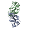
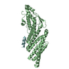
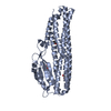
- PDB-1v1p: The structure SSL from Staphylococcus Aureus from an orthorhombic... -
+ Open data

- Basic information
| Entry | Database: PDB / ID: 1v1p | ||||||
|---|---|---|---|---|---|---|---|
| Title | The structure SSL from Staphylococcus Aureus from an orthorhombic crystal form | ||||||
 Components Components | (Exotoxin 1) x 2 | ||||||
 Keywords Keywords | VIRULENCE FACTOR / ANTIGEN PRESENTING CELL / SECRETED PROTEIN / STAPHYLOCOCCAL EXOTOXIN 1 / SET1 / SUPERANTIGEN / OB-FOLD / {BETA}-GRASP | ||||||
| Function / homology |  Function and homology information Function and homology information | ||||||
| Biological species |   Staphylococcus aureus (bacteria) Staphylococcus aureus (bacteria) | ||||||
| Method |  X-RAY DIFFRACTION / SYNCHROTRON / MOLECULAR REPLACEMENT / Resolution: 2.7 Å X-RAY DIFFRACTION / SYNCHROTRON / MOLECULAR REPLACEMENT / Resolution: 2.7 Å | ||||||
 Authors Authors | Briggs, D.C. / Naylor, C.E. | ||||||
 Citation Citation | Journal: Infect.Immun. / Year: 2004 Title: Structural Relationships and Cellular Tropism of Staphylococcal Superantigen-Like Proteins Authors: Al-Shangiti, A. / Naylor, C.E. / Nair, S. / Briggs, D.C. / Henderson, B. / Chain, B. | ||||||
| History |
|
- Structure visualization
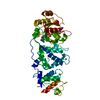
| Structure viewer | Molecule: MolmilJmol/JSmol |
|---|
- Downloads & links
-Download
| PDBx/mmCIF format | 1v1p.cif.gz | 88 KB | Display | PDBx/mmCIF format |
|---|---|---|---|---|
| PDB format | pdb1v1p.ent.gz | 66.2 KB | Display | PDB format |
| PDBx/mmJSON format | 1v1p.json.gz | Tree view | PDBx/mmJSON format | |
| Others |  Other downloads Other downloads |
-Validation report
| Arichive directory | https://data.pdbj.org/pub/pdb/validation_reports/v1/1v1pftp://data.pdbj.org/pub/pdb/validation_reports/v1/1v1p | HTTPS FTP |
|---|
-Related structure data
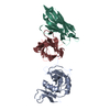
| Related structure data |  1v1oC 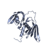 1m4vS C: citing same article ( S: Starting model for refinement |
|---|---|
| Similar structure data |


-Links
 PDBj
PDBj
- Assembly
| Deposited unit | 
| ||||||||
|---|---|---|---|---|---|---|---|---|---|
| 1 |
| ||||||||
| Unit cell |
| ||||||||
| Noncrystallographic symmetry (NCS) | NCS oper: (Code: given Matrix: (-0.999932, -0.011239, 0.003138), Vector: Details | THE ABOVE OLIGOMERIC STATE WAS DETERMINED FROM THEDEPOSITED COORDINATES USING THE PROTEIN QUATERNARYSTRUCTURE (PQS) ALGORITHM. HOWEVER, THE AUTHORS OF THISPDB ENTRY STRESS THAT THE BIOLOGICALLY SIGNIFICANT STATEFOR THIS ENTRY IS UNKNOWN AT THE TIME OF DEPOSITION. | |
-Components
| #1: Protein | Mass: 24234.094 Da / Num. of mol.: 1 Source method: isolated from a genetically manipulated source Source: (gene. exp.) Staphylococcus aureus (bacteria) / Gene: set1, ERS072840_02173, NCTC7878_00468 / Plasmid: PQE30 / Production host: |
|---|---|
| #2: Protein | Mass: 24478.357 Da / Num. of mol.: 1 Source method: isolated from a genetically manipulated source Source: (gene. exp.) Staphylococcus aureus (bacteria) / Gene: set1, ERS072840_02173, NCTC7878_00468 / Plasmid: PQE30 / Production host: |
| #3: Water | ChemComp-HOH /  Mass: 18.015 Da / Num. of mol.: 24 / Source method: isolated from a natural source / Formula: H2O Mass: 18.015 Da / Num. of mol.: 24 / Source method: isolated from a natural source / Formula: H2O |
| Sequence details | SIGNAL SEQUENCE REMOVED, HIS-TAG ADDED |
-Experimental details
-Experiment
| Experiment | Method: X-RAY DIFFRACTION / Number of used crystals: 1 |
|---|
- Sample preparation
| Crystal | Density Matthews: 2 Å3/Da / Density % sol: 65.2 % |
|---|---|
| Crystal grow | pH: 8.5 Details: 0.1M TRICINE BUFFER @PH 8.5, 28% (W/V) PEG2K, 0.1M LITHIUM SULPHATE |
-Data collection
| Diffraction | Mean temperature: 100 K |
|---|---|
| Diffraction source | Source: SYNCHROTRON / Site: ESRF  / Beamline: ID14-1 / Wavelength: 0.934 / Beamline: ID14-1 / Wavelength: 0.934 |
| Detector | Type: ADSC CCD / Detector: CCD / Date: Aug 15, 2004 / Details: GE(220) AND A MULTILAYER |
| Radiation | Monochromator: DIAMOND(111),GE(220) / Protocol: SINGLE WAVELENGTH / Monochromatic (M) / Laue (L): M / Scattering type: x-ray |
| Radiation wavelength | Wavelength: 0.934 Å / Relative weight: 1 |
| Reflection | Resolution: 2.7→31.01 Å / Num. obs: 11074 / % possible obs: 99.9 % / Redundancy: 4.5 % / Biso Wilson estimate: 55.92213 Å2 / Rmerge(I) obs: 0.14 / Net I/σ(I): 4.5902 |
| Reflection shell | Resolution: 2.7→2.85 Å / Redundancy: 3.9 % / Rmerge(I) obs: 0.49 / Mean I/σ(I) obs: 1.53 / % possible all: 99.9 |
- Processing
| Software |
| ||||||||||||||||||||
|---|---|---|---|---|---|---|---|---|---|---|---|---|---|---|---|---|---|---|---|---|---|
| Refinement | Method to determine structure: MOLECULAR REPLACEMENT Starting model: PDB ENTRY 1M4V Resolution: 2.7→58.722 Å / SU B: 17.44 / SU ML: 0.347 / Cross valid method: THROUGHOUT / ESU R Free: 0.447
| ||||||||||||||||||||
| Displacement parameters | Biso mean: 39.808 Å2
| ||||||||||||||||||||
| Refinement step | Cycle: LAST / Resolution: 2.7→58.722 Å
|
