 Movie
Movie Controller

[English] 日本語
 Yorodumi
- PDB-1v0f: Endosialidase of Bacteriophage K1F in complex with oligomeric alp... -
+ Open data

- Basic information
| Entry | Database: PDB / ID: 1v0f | |||||||||
|---|---|---|---|---|---|---|---|---|---|---|
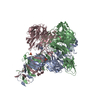
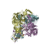
| Title | Endosialidase of Bacteriophage K1F in complex with oligomeric alpha-2,8-sialic acid | |||||||||
 Components Components | ENDO-ALPHA-SIALIDASE | |||||||||
 Keywords Keywords | HYDROLASE / ENDOSIALIDASE / POLYSIALIC ACID DEGRADATION / GLYCOSIDASE. | |||||||||
| Function / homology |  Function and homology information Function and homology informationendo-alpha-sialidase / endo-alpha-(2,8)-sialidase activity / symbiont entry into host cell via disruption of host cell glycocalyx / virus tail, fiber / symbiont entry into host cell via disruption of host cell envelope / symbiont entry into host / adhesion receptor-mediated virion attachment to host cell / virion attachment to host cell / identical protein binding Similarity search - Function | |||||||||
| Biological species |  COLIPHAGE K1F (virus) COLIPHAGE K1F (virus) | |||||||||
| Method |  X-RAY DIFFRACTION / SYNCHROTRON / OTHER / Resolution: 2.55 Å X-RAY DIFFRACTION / SYNCHROTRON / OTHER / Resolution: 2.55 Å | |||||||||
 Authors Authors | Stummeyer, K. / Dickmanns, A. / Muehlenhoff, M. / Gerady-Schahn, R. / Ficner, R. | |||||||||
 Citation Citation | Journal: Nat.Struct.Mol.Biol. / Year: 2005 Title: Crystal Structure of the Polysialic Acid-Degrading Endosialidase of Bacteriophage K1F Authors: Stummeyer, K. / Dickmanns, A. / Muehlenhoff, M. / Gerardy-Schahn, R. / Ficner, R. #1: Journal: J.Biol.Chem. / Year: 2003 Title: Proteolytic Processing and Oligomerization of Bacteriophage-Derived Endosialidases Authors: Muhlenhoff, M. / Stummeyer, K. / Grove, M. / Sauerborn, M. / Gerardy-Schahn, R. | |||||||||
| History |
| |||||||||
| Remark 700 | SHEET THE SHEET STRUCTURE OF THIS MOLECULE IS BIFURCATED. IN ORDER TO REPRESENT THIS FEATURE IN ... SHEET THE SHEET STRUCTURE OF THIS MOLECULE IS BIFURCATED. IN ORDER TO REPRESENT THIS FEATURE IN THE SHEET RECORDS BELOW, TWO SHEETS ARE DEFINED. |
- Structure visualization
| Structure viewer | Molecule: MolmilJmol/JSmol |
|---|
- Downloads & links
-Download
| PDBx/mmCIF format | 1v0f.cif.gz | 759.7 KB | Display | PDBx/mmCIF format |
|---|---|---|---|---|
| PDB format | pdb1v0f.ent.gz | 628.4 KB | Display | PDB format |
| PDBx/mmJSON format | 1v0f.json.gz | Tree view | PDBx/mmJSON format | |
| Others |  Other downloads Other downloads |
-Validation report
| Summary document | 1v0f_validation.pdf.gz | 3.2 MB | Display | wwPDB validaton report |
|---|---|---|---|---|
| Full document | 1v0f_full_validation.pdf.gz | 3.3 MB | Display | |
| Data in XML | 1v0f_validation.xml.gz | 149.6 KB | Display | |
| Data in CIF | 1v0f_validation.cif.gz | 204.4 KB | Display | |
| Arichive directory | https://data.pdbj.org/pub/pdb/validation_reports/v0/1v0fftp://data.pdbj.org/pub/pdb/validation_reports/v0/1v0f | HTTPS FTP |
-Related structure data


-Links
 PDBj
PDBj


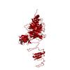
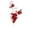
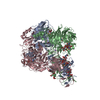
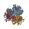
- Assembly
| Deposited unit | 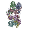
| ||||||||||||||||||||||||
|---|---|---|---|---|---|---|---|---|---|---|---|---|---|---|---|---|---|---|---|---|---|---|---|---|---|
| 1 | 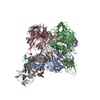
| ||||||||||||||||||||||||
| 2 | 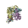
| ||||||||||||||||||||||||
| Unit cell |
| ||||||||||||||||||||||||
| Components on special symmetry positions |
| ||||||||||||||||||||||||
| Noncrystallographic symmetry (NCS) | NCS oper:
|
-Components
| #1: Protein | Mass: 74131.969 Da / Num. of mol.: 6 / Fragment: CATALYTIC DOMAIN, RESIDUES 246-911 Source method: isolated from a genetically manipulated source Source: (gene. exp.) COLIPHAGE K1F (virus) / Production host:  References: UniProt: Q858B1, UniProt: Q04830*PLUS, endo-alpha-sialidase #2: Polysaccharide | Source method: isolated from a genetically manipulated source #3: Sugar | ChemComp-SLB / 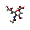  Type: D-saccharide, beta linking / Mass: 309.270 Da / Num. of mol.: 6 Type: D-saccharide, beta linking / Mass: 309.270 Da / Num. of mol.: 6Source method: isolated from a genetically manipulated source Formula: C11H19NO9 #4: Chemical | ChemComp-PO4 /   Mass: 94.971 Da / Num. of mol.: 6 / Source method: obtained synthetically / Formula: PO4 Mass: 94.971 Da / Num. of mol.: 6 / Source method: obtained synthetically / Formula: PO4#5: Water | ChemComp-HOH / |  Mass: 18.015 Da / Num. of mol.: 921 / Source method: isolated from a natural source / Formula: H2O Mass: 18.015 Da / Num. of mol.: 921 / Source method: isolated from a natural source / Formula: H2O |
|---|
-Experimental details
-Experiment
| Experiment | Method: X-RAY DIFFRACTION / Number of used crystals: 1 |
|---|
- Sample preparation
| Crystal | Density Matthews: 2.54 Å3/Da / Density % sol: 51.65 % |
|---|---|
| Crystal grow | pH: 7.5 / Details: pH 7.50 |
-Data collection
| Diffraction | Mean temperature: 100 K |
|---|---|
| Diffraction source | Source: SYNCHROTRON / Site: BESSY  / Beamline: 14.2 / Wavelength: 0.9195 / Beamline: 14.2 / Wavelength: 0.9195 |
| Detector | Type: MAR scanner 345 mm plate / Detector: IMAGE PLATE |
| Radiation | Protocol: SINGLE WAVELENGTH / Monochromatic (M) / Laue (L): M / Scattering type: x-ray |
| Radiation wavelength | Wavelength: 0.9195 Å / Relative weight: 1 |
| Reflection | Resolution: 2.55→30 Å / Num. obs: 130540 / % possible obs: 88.4 % / Redundancy: 4.3 % / Rmerge(I) obs: 0.107 / Net I/σ(I): 5.7 |
| Reflection shell | Resolution: 2.55→2.7 Å / Redundancy: 4.5 % / Rmerge(I) obs: 0.303 / Mean I/σ(I) obs: 2.4 / % possible all: 79.8 |
- Processing
| Software |
| ||||||||||||||||||||
|---|---|---|---|---|---|---|---|---|---|---|---|---|---|---|---|---|---|---|---|---|---|
| Refinement | Method to determine structure: OTHER / Resolution: 2.55→30 Å / SU B: 10.739 / SU ML: 0.231 / Cross valid method: THROUGHOUT / ESU R Free: 0.321
| ||||||||||||||||||||
| Displacement parameters | Biso mean: 21.406 Å2
| ||||||||||||||||||||
| Refinement step | Cycle: LAST / Resolution: 2.55→30 Å
|
