 Movie
Movie Controller

[English] 日本語
 Yorodumi
- PDB-1usy: ATP phosphoribosyl transferase (HisG:HisZ) complex from Thermotog... -
+ Open data

- Basic information
| Entry | Database: PDB / ID: 1usy | ||||||
|---|---|---|---|---|---|---|---|
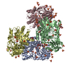
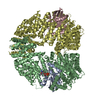
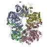
| Title | ATP phosphoribosyl transferase (HisG:HisZ) complex from Thermotoga maritima | ||||||
 Components Components |
| ||||||
 Keywords Keywords | TRANSFERASE / ATP PHOSPHORIBOSYL TRANSFERASE / AMINOACYL-TRNA SYNTHETASE | ||||||
| Function / homology |  Function and homology information Function and homology informationATP phosphoribosyltransferase / ATP phosphoribosyltransferase activity / histidine-tRNA ligase activity / histidyl-tRNA aminoacylation / L-histidine biosynthetic process / ATP binding / cytosol / cytoplasm Similarity search - Function | ||||||
| Biological species |   THERMOTOGA MARITIMA (bacteria) THERMOTOGA MARITIMA (bacteria) | ||||||
| Method |  X-RAY DIFFRACTION / SYNCHROTRON / MAD / Resolution: 2.52 Å X-RAY DIFFRACTION / SYNCHROTRON / MAD / Resolution: 2.52 Å | ||||||
 Authors Authors | Vega, M.C. / Fernandez, F.J. / Murphy, G.E. / Zou, P. / Popov, A. / Wilmanns, M. | ||||||
 Citation Citation | Journal: Mol.Microbiol. / Year: 2005 Title: Regulation of the Hetero-Octameric ATP Phosphoribosyl Transferase Complex from Thermotoga Maritima by a tRNA Synthetase-Like Subunit. Authors: Vega, M.C. / Zou, P. / Fernandez, F.J. / Murphy, G.E. / Sterner, R. / Popov, A. / Wilmanns, M. | ||||||
| History |
| ||||||
| Remark 700 | SHEET THE SHEET STRUCTURE OF THIS MOLECULE IS BIFURCATED. IN ORDER TO REPRESENT THIS FEATURE IN ... SHEET THE SHEET STRUCTURE OF THIS MOLECULE IS BIFURCATED. IN ORDER TO REPRESENT THIS FEATURE IN THE SHEET RECORDS BELOW, TWO SHEETS ARE DEFINED. |
- Structure visualization
| Structure viewer | Molecule: MolmilJmol/JSmol |
|---|
- Downloads & links
-Download
| PDBx/mmCIF format | 1usy.cif.gz | 391 KB | Display | PDBx/mmCIF format |
|---|---|---|---|---|
| PDB format | pdb1usy.ent.gz | 320 KB | Display | PDB format |
| PDBx/mmJSON format | 1usy.json.gz | Tree view | PDBx/mmJSON format | |
| Others |  Other downloads Other downloads |
-Validation report
| Arichive directory | https://data.pdbj.org/pub/pdb/validation_reports/us/1usyftp://data.pdbj.org/pub/pdb/validation_reports/us/1usy | HTTPS FTP |
|---|
-Related structure data
| Similar structure data |
|---|


-Links
 PDBj
PDBj

- Assembly
| Deposited unit | 
| ||||||||
|---|---|---|---|---|---|---|---|---|---|
| 1 |
| ||||||||
| Unit cell |
|
-Components
-ATP PHOSPHORIBOSYLTRANSFERASE REGULATORY ... , 2 types, 4 molecules ABDC
| #1: Protein | Mass: 31326.562 Da / Num. of mol.: 3 Source method: isolated from a genetically manipulated source Source: (gene. exp.) THERMOTOGA MARITIMA (bacteria) / Strain: MSB8 / Production host: #2: Protein | | Mass: 31354.617 Da / Num. of mol.: 1 Source method: isolated from a genetically manipulated source Source: (gene. exp.) THERMOTOGA MARITIMA (bacteria) / Strain: MSB8 / Production host: |
|---|
-Protein , 2 types, 4 molecules EFGH
| #3: Protein | Mass: 23544.416 Da / Num. of mol.: 3 Source method: isolated from a genetically manipulated source Source: (gene. exp.) THERMOTOGA MARITIMA (bacteria) / Strain: MSB8 / Production host: #4: Protein | | Mass: 23544.482 Da / Num. of mol.: 1 Source method: isolated from a genetically manipulated source Source: (gene. exp.) THERMOTOGA MARITIMA (bacteria) / Strain: MSB8 / Production host: |
|---|
-Non-polymers , 3 types, 288 molecules 


| #5: Chemical | ChemComp-HIS /  Type: L-peptide linking / Mass: 156.162 Da / Num. of mol.: 8 / Source method: obtained synthetically / Formula: C6H10N3O2 Type: L-peptide linking / Mass: 156.162 Da / Num. of mol.: 8 / Source method: obtained synthetically / Formula: C6H10N3O2#6: Chemical | ChemComp-PO4 /  Mass: 94.971 Da / Num. of mol.: 6 / Source method: obtained synthetically / Formula: PO4 Mass: 94.971 Da / Num. of mol.: 6 / Source method: obtained synthetically / Formula: PO4#7: Water | ChemComp-HOH / | Mass: 18.015 Da / Num. of mol.: 274 / Source method: isolated from a natural source / Formula: H2O |
|---|
-Details
| Has protein modification | Y |
|---|
-Experimental details
-Experiment
| Experiment | Method: X-RAY DIFFRACTION / Number of used crystals: 1 |
|---|
- Sample preparation
| Crystal | Density Matthews: 2.44 Å3/Da / Density % sol: 49.2 % |
|---|---|
| Crystal grow | pH: 4.2 / Details: pH 4.20 |
-Data collection
| Diffraction | Mean temperature: 100 K |
|---|---|
| Diffraction source | Source: SYNCHROTRON / Site: MPG/DESY, HAMBURG  / Beamline: BW6 / Wavelength: 0.9788 / Beamline: BW6 / Wavelength: 0.9788 |
| Detector | Type: MARRESEARCH / Detector: CCD / Date: Jun 15, 2002 / Details: COLLIMATOR |
| Radiation | Protocol: MAD / Monochromatic (M) / Laue (L): M / Scattering type: x-ray |
| Radiation wavelength | Wavelength: 0.9788 Å / Relative weight: 1 |
| Reflection | Resolution: 2.6→30 Å / Num. obs: 52743 / % possible obs: 98.4 % / Redundancy: 17.6 % / Rmerge(I) obs: 0.055 / Net I/σ(I): 18.44 |
| Reflection shell | Resolution: 2.6→2.7 Å / Redundancy: 4 % / Rmerge(I) obs: 0.289 / Mean I/σ(I) obs: 6.86 / % possible all: 98.1 |
- Processing
| Software |
| ||||||||||||||||||||||||||||||||||||||||||||||||||||||||||||||||||||||||||||||||||||||||||||||||||||||||||||||||||||||||||||||||||||||||||||||||||||||||||||||||||||||||||||||||||||||
|---|---|---|---|---|---|---|---|---|---|---|---|---|---|---|---|---|---|---|---|---|---|---|---|---|---|---|---|---|---|---|---|---|---|---|---|---|---|---|---|---|---|---|---|---|---|---|---|---|---|---|---|---|---|---|---|---|---|---|---|---|---|---|---|---|---|---|---|---|---|---|---|---|---|---|---|---|---|---|---|---|---|---|---|---|---|---|---|---|---|---|---|---|---|---|---|---|---|---|---|---|---|---|---|---|---|---|---|---|---|---|---|---|---|---|---|---|---|---|---|---|---|---|---|---|---|---|---|---|---|---|---|---|---|---|---|---|---|---|---|---|---|---|---|---|---|---|---|---|---|---|---|---|---|---|---|---|---|---|---|---|---|---|---|---|---|---|---|---|---|---|---|---|---|---|---|---|---|---|---|---|---|---|---|
| Refinement | Method to determine structure: MAD / Resolution: 2.52→12 Å / Cor.coef. Fo:Fc: 0.946 / Cor.coef. Fo:Fc free: 0.892 / SU B: 11.84 / SU ML: 0.263 / TLS residual ADP flag: LIKELY RESIDUAL / Cross valid method: THROUGHOUT / ESU R: 1.05 / ESU R Free: 0.359 / Stereochemistry target values: MAXIMUM LIKELIHOOD
| ||||||||||||||||||||||||||||||||||||||||||||||||||||||||||||||||||||||||||||||||||||||||||||||||||||||||||||||||||||||||||||||||||||||||||||||||||||||||||||||||||||||||||||||||||||||
| Solvent computation | Ion probe radii: 0.8 Å / Shrinkage radii: 0.8 Å / VDW probe radii: 1.4 Å / Solvent model: BABINET MODEL WITH MASK | ||||||||||||||||||||||||||||||||||||||||||||||||||||||||||||||||||||||||||||||||||||||||||||||||||||||||||||||||||||||||||||||||||||||||||||||||||||||||||||||||||||||||||||||||||||||
| Displacement parameters | Biso mean: 31.97 Å2
| ||||||||||||||||||||||||||||||||||||||||||||||||||||||||||||||||||||||||||||||||||||||||||||||||||||||||||||||||||||||||||||||||||||||||||||||||||||||||||||||||||||||||||||||||||||||
| Refinement step | Cycle: LAST / Resolution: 2.52→12 Å
| ||||||||||||||||||||||||||||||||||||||||||||||||||||||||||||||||||||||||||||||||||||||||||||||||||||||||||||||||||||||||||||||||||||||||||||||||||||||||||||||||||||||||||||||||||||||
| Refine LS restraints |
|