 Movie
Movie Controller

[English] 日本語
 Yorodumi
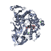
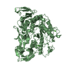
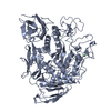
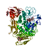
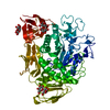
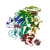
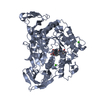
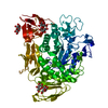
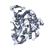
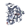
- PDB-1ukt: Crystal structure of Y100L mutant cyclodextrin glucanotransferase... -
+ Open data

- Basic information
| Entry | Database: PDB / ID: 1ukt | |||||||||
|---|---|---|---|---|---|---|---|---|---|---|
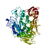
| Title | Crystal structure of Y100L mutant cyclodextrin glucanotransferase compexed with an acarbose | |||||||||
 Components Components | Cyclomaltodextrin glucanotransferase | |||||||||
 Keywords Keywords | TRANSFERASE / CGTASE / ACARBOSE / CARBOHYDRATE/PROTEIN INTERACTION | |||||||||
| Function / homology |  Function and homology information Function and homology informationcyclomaltodextrin glucanotransferase / cyclomaltodextrin glucanotransferase activity / starch binding / alpha-amylase activity / carbohydrate metabolic process / extracellular region / metal ion binding Similarity search - Function | |||||||||
| Biological species |  | |||||||||
| Method |  X-RAY DIFFRACTION / MOLECULAR REPLACEMENT / Resolution: 2.2 Å X-RAY DIFFRACTION / MOLECULAR REPLACEMENT / Resolution: 2.2 Å | |||||||||
 Authors Authors | Haga, K. / Kanai, R. / Sakamoto, O. / Harata, K. / Yamane, K. | |||||||||
 Citation Citation | Journal: J.Biochem.(Tokyo) / Year: 2003 Title: Effects of Essential Carbohydrate/Aromatic Stacking Interaction with Tyr100 and Phe259 on Substrate Binding of Cyclodextrin Glycosyltransferase from Alkalophilic Bacillus sp. 1011 Authors: Haga, K. / Kanai, R. / Sakamoto, O. / Aoyagi, M. / Harata, K. / Yamane, K. #1: Journal: Acta Crystallogr.,Sect.D / Year: 1996Title: X-ray Structure of Cyclodextrin Glucano-transferase from Alkalophilic Bacillus sp.1011. Comparison of Two Independent Molecules at 1.8 Angstrom Resolution Authors: Harata, K. / Haga, K. / Nakamura, A. / Aoyagi, M. / Yamane, K. | |||||||||
| History |
|
- Structure visualization
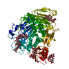
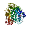
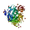
| Structure viewer | Molecule: MolmilJmol/JSmol |
|---|
- Downloads & links
-Download
| PDBx/mmCIF format | 1ukt.cif.gz | 278.4 KB | Display | PDBx/mmCIF format |
|---|---|---|---|---|
| PDB format | pdb1ukt.ent.gz | 222.8 KB | Display | PDB format |
| PDBx/mmJSON format | 1ukt.json.gz | Tree view | PDBx/mmJSON format | |
| Others |  Other downloads Other downloads |
-Validation report
| Arichive directory | https://data.pdbj.org/pub/pdb/validation_reports/uk/1uktftp://data.pdbj.org/pub/pdb/validation_reports/uk/1ukt | HTTPS FTP |
|---|
-Related structure data
| Related structure data |  1ukqC  1uksC 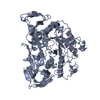 1pamS S: Starting model for refinement C: citing same article ( |
|---|---|
| Similar structure data |


-Links
 PDBj
PDBj


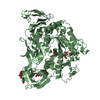
- Assembly
| Deposited unit | 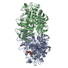
| ||||||||
|---|---|---|---|---|---|---|---|---|---|
| 1 | 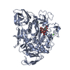
| ||||||||
| 2 | 
| ||||||||
| Unit cell |
|
-Components
| #1: Protein | Mass: 75180.070 Da / Num. of mol.: 2 / Mutation: Y100L Source method: isolated from a genetically manipulated source Source: (gene. exp.) References: UniProt: P05618, cyclomaltodextrin glucanotransferase #2: Polysaccharide | Source method: isolated from a genetically manipulated source #3: Chemical | 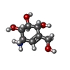  Mass: 175.182 Da / Num. of mol.: 2 Mass: 175.182 Da / Num. of mol.: 2Source method: isolated from a genetically manipulated source Formula: C7H13NO4 / Comment: antibiotic*YM #4: Chemical | ChemComp-CA /   Mass: 40.078 Da / Num. of mol.: 4 / Source method: obtained synthetically / Formula: Ca Mass: 40.078 Da / Num. of mol.: 4 / Source method: obtained synthetically / Formula: Ca#5: Water | ChemComp-HOH / |  Mass: 18.015 Da / Num. of mol.: 253 / Source method: isolated from a natural source / Formula: H2O Mass: 18.015 Da / Num. of mol.: 253 / Source method: isolated from a natural source / Formula: H2OHas protein modification | Y | |
|---|
-Experimental details
-Experiment
| Experiment | Method: X-RAY DIFFRACTION / Number of used crystals: 2 |
|---|
- Sample preparation
| Crystal | Density Matthews: 2.41 Å3/Da / Density % sol: 48.65 % |
|---|---|
| Crystal grow | Temperature: 293 K / Method: vapor diffusion, hanging drop / pH: 5.6 Details: PEG 3000, SODIUM CITRATE, 2-PROPANOL, CALCIUM CHLORIDE, ACARBOSE, pH 5.6, VAPOR DIFFUSION, HANGING DROP, temperature 293.0K |
| Crystal grow | *PLUS Method: unknown / Details: Harata, K., (1996) Acta Cryst., D52, 1136. |
-Data collection
| Diffraction | Mean temperature: 293 K |
|---|---|
| Diffraction source | Source: ROTATING ANODE / Type: RIGAKU / Wavelength: 1.5418 Å |
| Detector | Type: RIGAKU RAXIS IIC / Detector: IMAGE PLATE / Date: Feb 22, 1997 |
| Radiation | Monochromator: GRAPHITE / Protocol: SINGLE WAVELENGTH / Monochromatic (M) / Laue (L): M / Scattering type: x-ray |
| Radiation wavelength | Wavelength: 1.5418 Å / Relative weight: 1 |
| Reflection | Resolution: 2.01→76.77 Å / Num. all: 85791 / Num. obs: 68926 / Rmerge(I) obs: 0.068 |
| Reflection shell | Resolution: 2.01→2.05 Å / Rmerge(I) obs: 0.371 |
- Processing
| Software |
| ||||||||||||||||||||||||||||||||||||||||||
|---|---|---|---|---|---|---|---|---|---|---|---|---|---|---|---|---|---|---|---|---|---|---|---|---|---|---|---|---|---|---|---|---|---|---|---|---|---|---|---|---|---|---|---|
| Refinement | Method to determine structure: MOLECULAR REPLACEMENT Starting model: PDB ENTRY 1PAM Resolution: 2.2→10 Å / Isotropic thermal model: ISOTROPIC / σ(F): 2 / Stereochemistry target values: Engh & Huber
| ||||||||||||||||||||||||||||||||||||||||||
| Refinement step | Cycle: LAST / Resolution: 2.2→10 Å
| ||||||||||||||||||||||||||||||||||||||||||
| Refine LS restraints |
| ||||||||||||||||||||||||||||||||||||||||||
| LS refinement shell | Refine-ID: X-RAY DIFFRACTION / Total num. of bins used: 6
|
