 Movie
Movie Controller

[English] 日本語
 Yorodumi
- PDB-1ucr: Three-dimensional crystal structure of dissimilatory sulfite redu... -
+ Open data

- Basic information
| Entry | Database: PDB / ID: 1ucr | ||||||
|---|---|---|---|---|---|---|---|
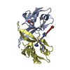
| Title | Three-dimensional crystal structure of dissimilatory sulfite reductase D (DsrD) | ||||||
 Components Components | Protein dsvD | ||||||
 Keywords Keywords | UNKNOWN FUNCTION / dissimilatory sulfite reductase D / DNA binding motif / sulfate-reducing bacteria / winged-helix motif | ||||||
| Function / homology | Dissimilatory sulphite reductase D / Dissimilatory sulfite reductase D (DsrD) / Winged helix-like DNA-binding domain superfamily/Winged helix DNA-binding domain / Arc Repressor Mutant, subunit A / Winged helix DNA-binding domain superfamily / Winged helix-like DNA-binding domain superfamily / Orthogonal Bundle / Mainly Alpha / Protein DsvD Function and homology information Function and homology information | ||||||
| Biological species |  Desulfovibrio vulgaris (bacteria) Desulfovibrio vulgaris (bacteria) | ||||||
| Method |  X-RAY DIFFRACTION / SYNCHROTRON / SIRAS / Resolution: 1.2 Å X-RAY DIFFRACTION / SYNCHROTRON / SIRAS / Resolution: 1.2 Å | ||||||
 Authors Authors | Mizuno, N. / Voordouw, G. / Miki, K. / Sarai, A. / Higuchi, Y. | ||||||
 Citation Citation | Journal: STRUCTURE / Year: 2003 Title: Crystal Structure of Dissimilatory Sulfite Reductase D (DsrD) Protein-Possible Interaction with B- and Z-DNA by Its Winged-Helix Motif Authors: Mizuno, N. / Voordouw, G. / Miki, K. / Sarai, A. / Higuchi, Y. | ||||||
| History |
|
- Structure visualization
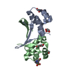
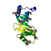
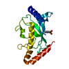
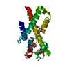
| Structure viewer | Molecule: MolmilJmol/JSmol |
|---|
- Downloads & links
-Download
| PDBx/mmCIF format | 1ucr.cif.gz | 82.7 KB | Display | PDBx/mmCIF format |
|---|---|---|---|---|
| PDB format | pdb1ucr.ent.gz | 62.8 KB | Display | PDB format |
| PDBx/mmJSON format | 1ucr.json.gz | Tree view | PDBx/mmJSON format | |
| Others |  Other downloads Other downloads |
-Validation report
| Arichive directory | https://data.pdbj.org/pub/pdb/validation_reports/uc/1ucrftp://data.pdbj.org/pub/pdb/validation_reports/uc/1ucr | HTTPS FTP |
|---|
-Related structure data
| Similar structure data |
|---|


-Links
 PDBj
PDBj- Assembly
| Deposited unit | 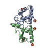
| ||||||||
|---|---|---|---|---|---|---|---|---|---|
| 1 |
| ||||||||
| Unit cell |
|
-Components
| #1: Protein | Mass: 8841.918 Da / Num. of mol.: 2 Source method: isolated from a genetically manipulated source Source: (gene. exp.) Desulfovibrio vulgaris (bacteria) / Production host: #2: Chemical | ChemComp-SO4 /   Mass: 96.063 Da / Num. of mol.: 5 / Source method: obtained synthetically / Formula: SO4 Mass: 96.063 Da / Num. of mol.: 5 / Source method: obtained synthetically / Formula: SO4#3: Water | ChemComp-HOH / |  Mass: 18.015 Da / Num. of mol.: 231 / Source method: isolated from a natural source / Formula: H2O Mass: 18.015 Da / Num. of mol.: 231 / Source method: isolated from a natural source / Formula: H2O |
|---|
-Experimental details
-Experiment
| Experiment | Method: X-RAY DIFFRACTION / Number of used crystals: 1 |
|---|
- Sample preparation
| Crystal | Density Matthews: 2.26 Å3/Da / Density % sol: 45.5 % | ||||||||||||||||||||
|---|---|---|---|---|---|---|---|---|---|---|---|---|---|---|---|---|---|---|---|---|---|
| Crystal grow | Temperature: 293 K / Method: vapor diffusion, sitting drop / pH: 5.3 Details: 73-75% saturated ammonium sulfate, pH 5.3, VAPOR DIFFUSION, SITTING DROP, temperature 293K | ||||||||||||||||||||
| Crystal grow | *PLUS Method: vapor diffusion, hanging drop / Details: Mizuno, N., (2000) Acta Crystallogr., D56, 754. | ||||||||||||||||||||
| Components of the solutions | *PLUS
|
-Data collection
| Diffraction | Mean temperature: 100 K |
|---|---|
| Diffraction source | Source: SYNCHROTRON / Site: SPring-8  / Beamline: BL44B2 / Wavelength: 0.708 Å / Beamline: BL44B2 / Wavelength: 0.708 Å |
| Detector | Type: MARRESEARCH / Detector: CCD / Date: Nov 2, 2000 |
| Radiation | Protocol: SINGLE WAVELENGTH / Monochromatic (M) / Laue (L): M / Scattering type: x-ray |
| Radiation wavelength | Wavelength: 0.708 Å / Relative weight: 1 |
| Reflection | Resolution: 1.2→100 Å / Num. obs: 55639 |
| Reflection | *PLUS Highest resolution: 1.2 Å / % possible obs: 99.8 % / Num. measured all: 565339 / Rmerge(I) obs: 0.06 |
| Reflection shell | *PLUS Highest resolution: 1.2 Å / Lowest resolution: 1.26 Å / % possible obs: 99.8 % / Num. unique obs: 8364 / Num. measured obs: 82965 / Rmerge(I) obs: 0.185 / Mean I/σ(I) obs: 1.1 |
- Processing
| Software |
| |||||||||||||||||||||||||||||||||
|---|---|---|---|---|---|---|---|---|---|---|---|---|---|---|---|---|---|---|---|---|---|---|---|---|---|---|---|---|---|---|---|---|---|---|
| Refinement | Method to determine structure: SIRAS / Resolution: 1.2→10 Å / Num. parameters: 13068 / Num. restraintsaints: 15368 / Cross valid method: THROUGHOUT / σ(F): 4 / Stereochemistry target values: Engh & Huber
| |||||||||||||||||||||||||||||||||
| Refine analyze | Num. disordered residues: 6 / Occupancy sum hydrogen: 0 / Occupancy sum non hydrogen: 1451 | |||||||||||||||||||||||||||||||||
| Refinement step | Cycle: LAST / Resolution: 1.2→10 Å
| |||||||||||||||||||||||||||||||||
| Refine LS restraints |
| |||||||||||||||||||||||||||||||||
| Software | *PLUS Name: SHELXL / Version: 97 / Classification: refinement | |||||||||||||||||||||||||||||||||
| Refinement | *PLUS Lowest resolution: 10 Å / Rfactor Rwork: 0.1389 | |||||||||||||||||||||||||||||||||
| Solvent computation | *PLUS | |||||||||||||||||||||||||||||||||
| Displacement parameters | *PLUS | |||||||||||||||||||||||||||||||||
| Refine LS restraints | *PLUS
| |||||||||||||||||||||||||||||||||
| LS refinement shell | *PLUS Highest resolution: 1.2 Å / Lowest resolution: 1.24 Å / Rfactor Rwork: 0.166 / Num. reflection obs: 5001 |
