 Movie
Movie Controller


+ Open data

- Basic information
| Entry | Database: PDB / ID: 1ucg | ||||||
|---|---|---|---|---|---|---|---|
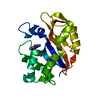
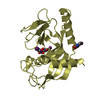
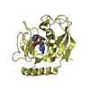
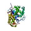
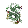
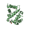
| Title | Crystal structure of Ribonuclease MC1 N71T mutant | ||||||
 Components Components | Ribonuclease MC | ||||||
 Keywords Keywords | HYDROLASE / alpha plus beta | ||||||
| Function / homology |  Function and homology information Function and homology informationribonuclease T2 / ribonuclease T2 activity / RNA catabolic process / hydrolase activity / RNA binding / extracellular region Similarity search - Function | ||||||
| Biological species |  Momordica charantia (bitter melon) Momordica charantia (bitter melon) | ||||||
| Method |  X-RAY DIFFRACTION / SYNCHROTRON / MOLECULAR REPLACEMENT / Resolution: 1.65 Å X-RAY DIFFRACTION / SYNCHROTRON / MOLECULAR REPLACEMENT / Resolution: 1.65 Å | ||||||
 Authors Authors | Suzuki, A. / Numata, T. / Yao, M. / Tanaka, I. / Kimura, M. | ||||||
 Citation Citation | Journal: Biochemistry / Year: 2003 Title: Crystal structures of the ribonuclease MC1 mutants N71T and N71S in complex with 5'-GMP: structural basis for alterations in substrate specificity Authors: Numata, T. / Suzuki, A. / Kakuta, Y. / Kimura, K. / Yao, M. / Tanaka, I. / Yoshida, Y. / Ueda, T. / Kimura, M. #1: Journal: Biochemistry / Year: 2001Title: Amino acid residues in ribonuclease MC1 from bitter gourd seeds which are essential for uridine specificity Authors: Numata, T. / Suzuki, A. / Yao, M. / Tanaka, I. / Kimura, M. | ||||||
| History |
|
- Structure visualization
| Structure viewer | Molecule: MolmilJmol/JSmol |
|---|
- Downloads & links
-Download
| PDBx/mmCIF format | 1ucg.cif.gz | 90.2 KB | Display | PDBx/mmCIF format |
|---|---|---|---|---|
| PDB format | pdb1ucg.ent.gz | 68.7 KB | Display | PDB format |
| PDBx/mmJSON format | 1ucg.json.gz | Tree view | PDBx/mmJSON format | |
| Others |  Other downloads Other downloads |
-Validation report
| Arichive directory | https://data.pdbj.org/pub/pdb/validation_reports/uc/1ucgftp://data.pdbj.org/pub/pdb/validation_reports/uc/1ucg | HTTPS FTP |
|---|
-Related structure data
| Related structure data |  1j1fC 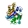 1j1gC  1bk7S S: Starting model for refinement C: citing same article ( |
|---|---|
| Similar structure data |


-Links
 PDBj
PDBj- Assembly
| Deposited unit | 
| ||||||||
|---|---|---|---|---|---|---|---|---|---|
| 1 | 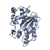
| ||||||||
| 2 | 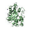
| ||||||||
| Unit cell |
| ||||||||
| Details | There are two monomers in the asymmetric unit, each monomer is a biological unit. |
-Components
| #1: Protein | Mass: 21198.033 Da / Num. of mol.: 2 / Mutation: N71T Source method: isolated from a genetically manipulated source Source: (gene. exp.) Momordica charantia (bitter melon) / Plasmid: pET-22b / Production host:  #2: Chemical | ChemComp-MN /   Mass: 54.938 Da / Num. of mol.: 4 / Source method: obtained synthetically / Formula: Mn Mass: 54.938 Da / Num. of mol.: 4 / Source method: obtained synthetically / Formula: Mn#3: Water | ChemComp-HOH / |  Mass: 18.015 Da / Num. of mol.: 231 / Source method: isolated from a natural source / Formula: H2O Mass: 18.015 Da / Num. of mol.: 231 / Source method: isolated from a natural source / Formula: H2OHas protein modification | Y | |
|---|
-Experimental details
-Experiment
| Experiment | Method: X-RAY DIFFRACTION / Number of used crystals: 1 |
|---|
- Sample preparation
| Crystal | Density Matthews: 2.47 Å3/Da / Density % sol: 50.14 % | ||||||||||||||||||||||||
|---|---|---|---|---|---|---|---|---|---|---|---|---|---|---|---|---|---|---|---|---|---|---|---|---|---|
| Crystal grow | Temperature: 293 K / Method: vapor diffusion, hanging drop / pH: 7 Details: PEG 1540, Manganese Cloride tetrahydrate, pH 7.0, VAPOR DIFFUSION, HANGING DROP, temperature 293K | ||||||||||||||||||||||||
| Crystal grow | *PLUS Temperature: 20 ℃ | ||||||||||||||||||||||||
| Components of the solutions | *PLUS
|
-Data collection
| Diffraction | Mean temperature: 100 K |
|---|---|
| Diffraction source | Source: SYNCHROTRON / Site: SPring-8  / Beamline: BL40B2 / Wavelength: 1 Å / Beamline: BL40B2 / Wavelength: 1 Å |
| Detector | Type: ADSC QUANTUM 4 / Detector: CCD / Date: May 25, 2001 / Details: mirrors |
| Radiation | Monochromator: Mirror / Protocol: SINGLE WAVELENGTH / Monochromatic (M) / Laue (L): M / Scattering type: x-ray |
| Radiation wavelength | Wavelength: 1 Å / Relative weight: 1 |
| Reflection | Resolution: 1.65→19.96 Å / Num. all: 50912 / Num. obs: 50884 / % possible obs: 99.2 % / Observed criterion σ(I): 3 / Redundancy: 7.6 % / Biso Wilson estimate: 36.31 Å2 / Rmerge(I) obs: 0.053 / Rsym value: 0.05 / Net I/σ(I): 9.1 |
| Reflection shell | Resolution: 1.65→1.74 Å / Redundancy: 4.8 % / Rmerge(I) obs: 0.212 / Mean I/σ(I) obs: 3.8 / Num. unique all: 7159 / Rsym value: 0.189 / % possible all: 97.3 |
| Reflection | *PLUS Lowest resolution: 35.81 Å / Num. obs: 50905 / Redundancy: 8.5 % / Num. measured all: 434769 |
| Reflection shell | *PLUS % possible obs: 97.6 % / Redundancy: 5.7 % / Rmerge(I) obs: 0.24 / Mean I/σ(I) obs: 22.3 |
- Processing
| Software |
| |||||||||||||||||||||||||
|---|---|---|---|---|---|---|---|---|---|---|---|---|---|---|---|---|---|---|---|---|---|---|---|---|---|---|
| Refinement | Method to determine structure: MOLECULAR REPLACEMENT Starting model: PDB ENTRY 1BK7 Resolution: 1.65→10 Å / Cross valid method: THROUGHOUT / σ(F): 0 / Stereochemistry target values: Engh & Huber
| |||||||||||||||||||||||||
| Displacement parameters | Biso mean: 23.94 Å2
| |||||||||||||||||||||||||
| Refine analyze |
| |||||||||||||||||||||||||
| Refinement step | Cycle: LAST / Resolution: 1.65→10 Å
| |||||||||||||||||||||||||
| Refine LS restraints |
| |||||||||||||||||||||||||
| LS refinement shell | Resolution: 1.65→1.71 Å
| |||||||||||||||||||||||||
| Refinement | *PLUS % reflection Rfree: 5 % / Rfactor Rfree: 0.195 | |||||||||||||||||||||||||
| Solvent computation | *PLUS | |||||||||||||||||||||||||
| Displacement parameters | *PLUS | |||||||||||||||||||||||||
| Refine LS restraints | *PLUS
|
