 Movie
Movie Controller

[English] 日本語
 Yorodumi
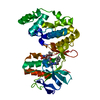
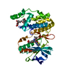
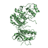
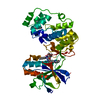
- PDB-1ubd: CO-CRYSTAL STRUCTURE OF HUMAN YY1 ZINC FINGER DOMAIN BOUND TO THE... -
+ Open data

- Basic information
| Entry | Database: PDB / ID: 1ubd | ||||||
|---|---|---|---|---|---|---|---|
| Title | CO-CRYSTAL STRUCTURE OF HUMAN YY1 ZINC FINGER DOMAIN BOUND TO THE ADENO-ASSOCIATED VIRUS P5 INITIATOR ELEMENT | ||||||
 Components Components |
| ||||||
 Keywords Keywords | TRANSCRIPTION/DNA / TRANSCRIPTION INITIATION / INITIATOR ELEMENT / YY1 / ZINC FINGER PROTEIN / DNA- PROTEIN RECOGNITION / COMPLEX (TRANSCRIPTION REGULATION-DNA) / TRANSCRIPTION-DNA COMPLEX | ||||||
| Function / homology |  Function and homology information Function and homology informationresponse to prostaglandin F / regulation of DNA strand elongation / positive regulation of telomere maintenance in response to DNA damage / negative regulation of cell growth involved in cardiac muscle cell development / PcG protein complex / negative regulation of interferon-beta production / Ino80 complex / chromatin silencing complex / response to UV-C / DNA-binding transcription repressor activity ...response to prostaglandin F / regulation of DNA strand elongation / positive regulation of telomere maintenance in response to DNA damage / negative regulation of cell growth involved in cardiac muscle cell development / PcG protein complex / negative regulation of interferon-beta production / Ino80 complex / chromatin silencing complex / response to UV-C / DNA-binding transcription repressor activity / immunoglobulin heavy chain V-D-J recombination / lncRNA binding / regulation of chromosome organization / SMAD binding / regulation of DNA replication / regulation of embryonic development / cellular response to interleukin-1 / cis-regulatory region sequence-specific DNA binding / four-way junction DNA binding / TFAP2 (AP-2) family regulates transcription of growth factors and their receptors / negative regulation of miRNA transcription / B cell differentiation / telomere maintenance / positive regulation of DNA repair / promoter-specific chromatin binding / DNA-binding transcription repressor activity, RNA polymerase II-specific / double-strand break repair via homologous recombination / DNA Damage Recognition in GG-NER / Activation of anterior HOX genes in hindbrain development during early embryogenesis / fibrillar center / nuclear matrix / sequence-specific double-stranded DNA binding / cellular response to UV / UCH proteinases / DNA-binding transcription activator activity, RNA polymerase II-specific / spermatogenesis / transcription regulator complex / Estrogen-dependent gene expression / DNA-binding transcription factor binding / DNA-binding transcription factor activity, RNA polymerase II-specific / regulation of cell cycle / transcription cis-regulatory region binding / nuclear body / RNA polymerase II cis-regulatory region sequence-specific DNA binding / chromatin remodeling / ribonucleoprotein complex / negative regulation of gene expression / DNA damage response / regulation of transcription by RNA polymerase II / positive regulation of DNA-templated transcription / chromatin / DNA-templated transcription / negative regulation of transcription by RNA polymerase II / positive regulation of transcription by RNA polymerase II / DNA binding / RNA binding / zinc ion binding / nucleoplasm / nucleus Similarity search - Function | ||||||
| Biological species |  Homo sapiens (human) Homo sapiens (human) | ||||||
| Method |  X-RAY DIFFRACTION / SYNCHROTRON / Resolution: 2.5 Å X-RAY DIFFRACTION / SYNCHROTRON / Resolution: 2.5 Å | ||||||
 Authors Authors | Houbaviy, H.B. / Usheva, A. / Shenk, T. / Burley, S.K. | ||||||
 Citation Citation | Journal: Proc.Natl.Acad.Sci.USA / Year: 1996 Title: Cocrystal structure of YY1 bound to the adeno-associated virus P5 initiator. Authors: Houbaviy, H.B. / Usheva, A. / Shenk, T. / Burley, S.K. | ||||||
| History |
|
- Structure visualization
| Structure viewer | Molecule: MolmilJmol/JSmol |
|---|
- Downloads & links
-Download
| PDBx/mmCIF format | 1ubd.cif.gz | 63.2 KB | Display | PDBx/mmCIF format |
|---|---|---|---|---|
| PDB format | pdb1ubd.ent.gz | 42.4 KB | Display | PDB format |
| PDBx/mmJSON format | 1ubd.json.gz | Tree view | PDBx/mmJSON format | |
| Others |  Other downloads Other downloads |
-Validation report
| Arichive directory | https://data.pdbj.org/pub/pdb/validation_reports/ub/1ubdftp://data.pdbj.org/pub/pdb/validation_reports/ub/1ubd | HTTPS FTP |
|---|
-Related structure data
| Similar structure data |
|---|


-Links
 PDBj
PDBj


- Assembly
| Deposited unit | 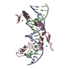
| ||||||||
|---|---|---|---|---|---|---|---|---|---|
| 1 |
| ||||||||
| Unit cell |
|
-Components
| #1: DNA chain | Mass: 6164.989 Da / Num. of mol.: 1 / Source method: obtained synthetically | ||
|---|---|---|---|
| #2: DNA chain | Mass: 6102.968 Da / Num. of mol.: 1 / Source method: obtained synthetically | ||
| #3: Protein | Mass: 14189.473 Da / Num. of mol.: 1 Source method: isolated from a genetically manipulated source Source: (gene. exp.) Homo sapiens (human) / Production host:  Keywords: MUTANT D291M, A292E / References: UniProt: P25490 Keywords: MUTANT D291M, A292E / References: UniProt: P25490 | ||
| #4: Chemical | ChemComp-ZN /   Mass: 65.409 Da / Num. of mol.: 4 / Source method: obtained synthetically / Formula: Zn Mass: 65.409 Da / Num. of mol.: 4 / Source method: obtained synthetically / Formula: Zn#5: Water | ChemComp-HOH / |  Mass: 18.015 Da / Num. of mol.: 87 / Source method: isolated from a natural source / Formula: H2O Mass: 18.015 Da / Num. of mol.: 87 / Source method: isolated from a natural source / Formula: H2O |
-Experimental details
-Experiment
| Experiment | Method: X-RAY DIFFRACTION |
|---|
- Sample preparation
| Crystal | Density Matthews: 3.14 Å3/Da / Density % sol: 60.84 % | ||||||||||||||||||||||||||||||||||||
|---|---|---|---|---|---|---|---|---|---|---|---|---|---|---|---|---|---|---|---|---|---|---|---|---|---|---|---|---|---|---|---|---|---|---|---|---|---|
| Crystal grow | *PLUS Temperature: 4 ℃ / Method: vapor diffusion | ||||||||||||||||||||||||||||||||||||
| Components of the solutions | *PLUS
|
-Data collection
| Diffraction source | Source: SYNCHROTRON / Site: NSLS  / Beamline: X12B / Beamline: X12B |
|---|---|
| Detector | Type: MARRESEARCH / Detector: IMAGE PLATE / Date: Jun 16, 1995 |
| Radiation | Protocol: SINGLE WAVELENGTH / Monochromatic (M) / Laue (L): M / Scattering type: x-ray |
| Radiation wavelength | Relative weight: 1 |
| Reflection | Num. obs: 12347 / % possible obs: 93.6 % / Observed criterion σ(I): 3 / Redundancy: 5 % / Rmerge(I) obs: 0.08 |
| Reflection | *PLUS Highest resolution: 2.5 Å / % possible obs: 93.6 % / Num. measured all: 64023 |
- Processing
| Software |
| ||||||||||||||||||||||||||||||||||||||||||||||||||||||||||||||||||||||||||||||||
|---|---|---|---|---|---|---|---|---|---|---|---|---|---|---|---|---|---|---|---|---|---|---|---|---|---|---|---|---|---|---|---|---|---|---|---|---|---|---|---|---|---|---|---|---|---|---|---|---|---|---|---|---|---|---|---|---|---|---|---|---|---|---|---|---|---|---|---|---|---|---|---|---|---|---|---|---|---|---|---|---|---|
| Refinement | Resolution: 2.5→6 Å / σ(F): 2 Details: LOCAL SCALING OF F(OBSERVED) AGAINST F(CALCULATED) WITH LSCALE (AUTHOR: M. ROULD) TO CORRECT FOR ANISOTROPIC DIFFRACTION AND ABSORPTION EFFECTS
| ||||||||||||||||||||||||||||||||||||||||||||||||||||||||||||||||||||||||||||||||
| Refinement step | Cycle: LAST / Resolution: 2.5→6 Å
| ||||||||||||||||||||||||||||||||||||||||||||||||||||||||||||||||||||||||||||||||
| Refine LS restraints |
| ||||||||||||||||||||||||||||||||||||||||||||||||||||||||||||||||||||||||||||||||
| Software | *PLUS Name: X-PLOR / Version: 3.1 / Classification: refinement | ||||||||||||||||||||||||||||||||||||||||||||||||||||||||||||||||||||||||||||||||
| Refinement | *PLUS Highest resolution: 2.5 Å / Lowest resolution: 6 Å / σ(F): 2 / Num. reflection Rfree: 1121 / Rfactor Rfree: 0.33 | ||||||||||||||||||||||||||||||||||||||||||||||||||||||||||||||||||||||||||||||||
| Solvent computation | *PLUS | ||||||||||||||||||||||||||||||||||||||||||||||||||||||||||||||||||||||||||||||||
| Displacement parameters | *PLUS | ||||||||||||||||||||||||||||||||||||||||||||||||||||||||||||||||||||||||||||||||
| Refine LS restraints | *PLUS
|
