 Movie
Movie Controller

[English] 日本語
 Yorodumi
- PDB-1ub9: Structure of the transcriptional regulator homologue protein from... -
+ Open data

- Basic information
| Entry | Database: PDB / ID: 1ub9 | ||||||
|---|---|---|---|---|---|---|---|
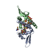
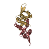
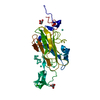
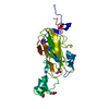
| Title | Structure of the transcriptional regulator homologue protein from Pyrococcus horikoshii OT3 | ||||||
 Components Components | Hypothetical protein PH1061 | ||||||
 Keywords Keywords | TRANSCRIPTION / helix-turn-helix motif / winged helix motif / STRUCTURAL GENOMICS | ||||||
| Function / homology |  Function and homology information Function and homology information | ||||||
| Biological species |   Pyrococcus horikoshii (archaea) Pyrococcus horikoshii (archaea) | ||||||
| Method |  X-RAY DIFFRACTION / SYNCHROTRON / MAD / Resolution: 2.05 Å X-RAY DIFFRACTION / SYNCHROTRON / MAD / Resolution: 2.05 Å | ||||||
 Authors Authors | Okada, U. / Sakai, N. / Tajika, Y. / Yao, M. / Watanabe, N. / Tanaka, I. | ||||||
 Citation Citation | Journal: Proteins / Year: 2006 Title: Structural analysis of the transcriptional regulator homolog protein from Pyrococcus horikoshii OT3. Authors: Okada, U. / Sakai, N. / Yao, M. / Watanabe, N. / Tanaka, I. | ||||||
| History |
|
- Structure visualization
| Structure viewer | Molecule: MolmilJmol/JSmol |
|---|
- Downloads & links
-Download
| PDBx/mmCIF format | 1ub9.cif.gz | 32 KB | Display | PDBx/mmCIF format |
|---|---|---|---|---|
| PDB format | pdb1ub9.ent.gz | 22 KB | Display | PDB format |
| PDBx/mmJSON format | 1ub9.json.gz | Tree view | PDBx/mmJSON format | |
| Others |  Other downloads Other downloads |
-Validation report
| Arichive directory | https://data.pdbj.org/pub/pdb/validation_reports/ub/1ub9ftp://data.pdbj.org/pub/pdb/validation_reports/ub/1ub9 | HTTPS FTP |
|---|
-Related structure data
| Similar structure data |
|---|


-Links
 PDBj
PDBj- Assembly
| Deposited unit | 
| ||||||||
|---|---|---|---|---|---|---|---|---|---|
| 1 | 
| ||||||||
| Unit cell |
| ||||||||
| Components on special symmetry positions |
| ||||||||
| Details | The biological assembly is a dimer by the operation: y, x,-z. |
-Components
| #1: Protein | Mass: 11402.514 Da / Num. of mol.: 1 Source method: isolated from a genetically manipulated source Source: (gene. exp.) Pyrococcus horikoshii (archaea) / Strain: OT3 / Gene: PH1061 / Plasmid: pET22b / Production host:  |
|---|---|
| #2: Water | ChemComp-HOH /  Mass: 18.015 Da / Num. of mol.: 57 / Source method: isolated from a natural source / Formula: H2O Mass: 18.015 Da / Num. of mol.: 57 / Source method: isolated from a natural source / Formula: H2O |
-Experimental details
-Experiment
| Experiment | Method: X-RAY DIFFRACTION / Number of used crystals: 1 |
|---|
- Sample preparation
| Crystal | Density Matthews: 1.94 Å3/Da / Density % sol: 43 % |
|---|---|
| Crystal grow | Temperature: 293 K / Method: vapor diffusion, hanging drop / pH: 8.3 Details: ammonium phosphate, pH 8.3, VAPOR DIFFUSION, HANGING DROP, temperature 293.0K |
-Data collection
| Diffraction | Mean temperature: 100 K | ||||||||||||
|---|---|---|---|---|---|---|---|---|---|---|---|---|---|
| Diffraction source | Source: SYNCHROTRON / Site: SPring-8  / Beamline: BL41XU / Wavelength: 0.9000,0.9794,0.9796 / Beamline: BL41XU / Wavelength: 0.9000,0.9794,0.9796 | ||||||||||||
| Detector | Type: MARRESEARCH / Detector: CCD / Date: Feb 8, 2003 / Details: mirrors | ||||||||||||
| Radiation | Monochromator: mirrors / Protocol: MAD / Monochromatic (M) / Laue (L): M / Scattering type: x-ray | ||||||||||||
| Radiation wavelength |
| ||||||||||||
| Reflection | Resolution: 2.05→33 Å / Num. all: 6696 / Num. obs: 6696 / % possible obs: 99.5 % / Observed criterion σ(I): 0 / Redundancy: 13.7 % / Biso Wilson estimate: 45.1 Å2 / Rmerge(I) obs: 0.06 / Net I/σ(I): 13.8 | ||||||||||||
| Reflection shell | Resolution: 2.05→2.12 Å / Redundancy: 14 % / Rmerge(I) obs: 0.234 / Num. unique all: 635 / % possible all: 100 |
- Processing
| Software |
| |||||||||||||||||||||||||||
|---|---|---|---|---|---|---|---|---|---|---|---|---|---|---|---|---|---|---|---|---|---|---|---|---|---|---|---|---|
| Refinement | Method to determine structure: MAD / Resolution: 2.05→10 Å / Isotropic thermal model: isotropic / Cross valid method: THROUGHOUT / σ(F): 0 / σ(I): 0 / Stereochemistry target values: Engh & Huber
| |||||||||||||||||||||||||||
| Solvent computation | Bsol: 55.8 Å2 / ksol: 0.3944 e/Å3 | |||||||||||||||||||||||||||
| Displacement parameters | Biso mean: 39.37 Å2
| |||||||||||||||||||||||||||
| Refine analyze |
| |||||||||||||||||||||||||||
| Refinement step | Cycle: LAST / Resolution: 2.05→10 Å
| |||||||||||||||||||||||||||
| Refine LS restraints |
| |||||||||||||||||||||||||||
| LS refinement shell | Resolution: 2.05→2.12 Å / Total num. of bins used: 10
| |||||||||||||||||||||||||||
| Xplor file | Serial no: 1 / Param file: protein_rep.param / Topol file: protein.top |
