 Movie
Movie Controller

[English] 日本語
 Yorodumi
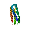
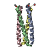
- PDB-1u9h: Heterocyclic Peptide Backbone Modification in GCN4-pLI Based Coil... -
+ Open data

- Basic information
| Entry | Database: PDB / ID: 1u9h | ||||||
|---|---|---|---|---|---|---|---|
| Title | Heterocyclic Peptide Backbone Modification in GCN4-pLI Based Coiled Coils: Replacement of E(22)L(23) | ||||||
 Components Components | General control protein GCN4 | ||||||
 Keywords Keywords | TRANSCRIPTION / tetrameric alpha-helical coiled coil / heterocycic backbone modification | ||||||
| Method |  X-RAY DIFFRACTION / MOLECULAR REPLACEMENT / Resolution: 2.17 Å X-RAY DIFFRACTION / MOLECULAR REPLACEMENT / Resolution: 2.17 Å | ||||||
 Authors Authors | Horne, W.S. / Yadav, M.K. / Stout, C.D. / Ghadiri, M.R. | ||||||
 Citation Citation | Journal: J.Am.Chem.Soc. / Year: 2004 Title: Heterocyclic peptide backbone modifications in an alpha-helical coiled coil. Authors: Horne, W.S. / Yadav, M.K. / Stout, C.D. / Ghadiri, M.R. | ||||||
| History |
|
- Structure visualization
| Structure viewer | Molecule:  MolmilJmol/JSmol MolmilJmol/JSmol |
|---|
- Downloads & links
-Download
| PDBx/mmCIF format | 1u9h.cif.gz | 25.8 KB | Display | PDBx/mmCIF format |
|---|---|---|---|---|
| PDB format | pdb1u9h.ent.gz | 17.6 KB | Display | PDB format |
| PDBx/mmJSON format | 1u9h.json.gz | Tree view | PDBx/mmJSON format | |
| Others |  Other downloads Other downloads |
-Validation report
| Arichive directory | https://data.pdbj.org/pub/pdb/validation_reports/u9/1u9hftp://data.pdbj.org/pub/pdb/validation_reports/u9/1u9h | HTTPS FTP |
|---|
-Related structure data


-Links
 PDBj
PDBj- Assembly
| Deposited unit | 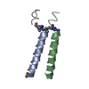
| |||||||||
|---|---|---|---|---|---|---|---|---|---|---|
| 1 | 
| |||||||||
| 2 |
| |||||||||
| Unit cell |
| |||||||||
| Components on special symmetry positions |
| |||||||||
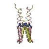
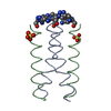
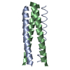
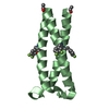
| Details | The tetramer formed by chains A and B is generated by the two fold axis: y,-x,1/4+z |
-Components
| #1: Protein/peptide | Mass: 4024.800 Da / Num. of mol.: 2 / Mutation: (TA4)22E,A23L / Source method: obtained synthetically Details: Prepared by Fmoc solid phase peptide synthesis. The sequence of this protein can be found naturally in Saccharomyces cerevisiae (Baker's yeast). #2: Water | ChemComp-HOH / |  Mass: 18.015 Da / Num. of mol.: 45 / Source method: isolated from a natural source / Formula: H2O Mass: 18.015 Da / Num. of mol.: 45 / Source method: isolated from a natural source / Formula: H2OHas protein modification | Y | |
|---|
-Experimental details
-Experiment
| Experiment | Method: X-RAY DIFFRACTION / Number of used crystals: 1 |
|---|
- Sample preparation
| Crystal | Density Matthews: 2.91 Å3/Da / Density % sol: 57.75 % |
|---|---|
| Crystal grow | Temperature: 295 K / Method: vapor diffusion, hanging drop / pH: 6.5 Details: 0.16 M magnesium acetate tetrahydrate, 0.08 M sodium cacodylate, pH 6.5, 16% w/v PEG 8000, 20% v/v anhydrous glycerol, VAPOR DIFFUSION, HANGING DROP, temperature 295K |
-Data collection
| Diffraction | Mean temperature: 100 K |
|---|---|
| Diffraction source | Source: ROTATING ANODE / Type: RIGAKU RU200 / Wavelength: 1.5418 Å |
| Detector | Type: RIGAKU RAXIS IV / Detector: IMAGE PLATE / Date: Apr 8, 2004 / Details: osmic confocal mirrors |
| Radiation | Protocol: SINGLE WAVELENGTH / Monochromatic (M) / Laue (L): M / Scattering type: x-ray |
| Radiation wavelength | Wavelength: 1.5418 Å / Relative weight: 1 |
| Reflection | Resolution: 2.17→45.17 Å / Num. obs: 5435 / % possible obs: 98.9 % / Observed criterion σ(I): 2 / Redundancy: 6.2 % / Rmerge(I) obs: 0.052 / Net I/σ(I): 7.2 |
| Reflection shell | Resolution: 2.17→2.25 Å / Rmerge(I) obs: 0.31 / Mean I/σ(I) obs: 2.4 / % possible all: 95.7 |
- Processing
| Software |
| ||||||||||||||||||||||||||||||||||||||||||||||||||||||||||||||||||||||||||||||||||||||||||||||||||||||||||||||||||||||||||||||||||||||||||||||||||||||||||||||||
|---|---|---|---|---|---|---|---|---|---|---|---|---|---|---|---|---|---|---|---|---|---|---|---|---|---|---|---|---|---|---|---|---|---|---|---|---|---|---|---|---|---|---|---|---|---|---|---|---|---|---|---|---|---|---|---|---|---|---|---|---|---|---|---|---|---|---|---|---|---|---|---|---|---|---|---|---|---|---|---|---|---|---|---|---|---|---|---|---|---|---|---|---|---|---|---|---|---|---|---|---|---|---|---|---|---|---|---|---|---|---|---|---|---|---|---|---|---|---|---|---|---|---|---|---|---|---|---|---|---|---|---|---|---|---|---|---|---|---|---|---|---|---|---|---|---|---|---|---|---|---|---|---|---|---|---|---|---|---|---|---|---|
| Refinement | Method to determine structure: MOLECULAR REPLACEMENT / Resolution: 2.17→45.17 Å / Cor.coef. Fo:Fc: 0.921 / Cor.coef. Fo:Fc free: 0.92 / SU B: 6.418 / SU ML: 0.162 / Cross valid method: THROUGHOUT / ESU R: 0.242 / ESU R Free: 0.209 / Stereochemistry target values: MAXIMUM LIKELIHOOD / Details: HYDROGENS HAVE BEEN ADDED IN THE RIDING POSITIONS
| ||||||||||||||||||||||||||||||||||||||||||||||||||||||||||||||||||||||||||||||||||||||||||||||||||||||||||||||||||||||||||||||||||||||||||||||||||||||||||||||||
| Solvent computation | Ion probe radii: 0.8 Å / Shrinkage radii: 0.8 Å / VDW probe radii: 1.2 Å / Solvent model: MASK | ||||||||||||||||||||||||||||||||||||||||||||||||||||||||||||||||||||||||||||||||||||||||||||||||||||||||||||||||||||||||||||||||||||||||||||||||||||||||||||||||
| Displacement parameters | Biso mean: 36.136 Å2
| ||||||||||||||||||||||||||||||||||||||||||||||||||||||||||||||||||||||||||||||||||||||||||||||||||||||||||||||||||||||||||||||||||||||||||||||||||||||||||||||||
| Refinement step | Cycle: LAST / Resolution: 2.17→45.17 Å
| ||||||||||||||||||||||||||||||||||||||||||||||||||||||||||||||||||||||||||||||||||||||||||||||||||||||||||||||||||||||||||||||||||||||||||||||||||||||||||||||||
| Refine LS restraints |
| ||||||||||||||||||||||||||||||||||||||||||||||||||||||||||||||||||||||||||||||||||||||||||||||||||||||||||||||||||||||||||||||||||||||||||||||||||||||||||||||||
| LS refinement shell | Resolution: 2.17→2.226 Å / Total num. of bins used: 20
|
