- PDB-1u3m: NMR structure of the chicken prion protein fragment 128-242 -
+
Open data
ID or keywords:
Loading...
-
Basic information
Entry
Database: PDB / ID: 1u3m
Title
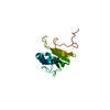
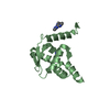
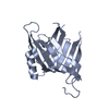
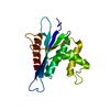
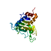
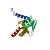
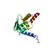
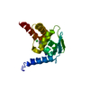
NMR structure of the chicken prion protein fragment 128-242
Components
prion-like protein
Keywords
MEMBRANE PROTEIN / PRION PROTEIN / TSE / PRP
Function / homology
Function and homology information
Insertion of tail-anchored proteins into the endoplasmic reticulum membrane / Post-translational modification: synthesis of GPI-anchored proteins / basement membrane / side of membrane / synaptic cleft / protein homooligomerization / copper ion binding / plasma membrane Similarity search - Function
Prion/Doppel protein, beta-ribbon domain / Major Prion Protein / Prion protein signature 1. / Prion protein signature 2. / Prion protein / Major prion protein / Prion/Doppel protein, beta-ribbon domain / Prion/Doppel beta-ribbon domain superfamily / Prion/Doppel alpha-helical domain / Orthogonal Bundle / Mainly Alpha Similarity search - Domain/homology
Protocol: SINGLE WAVELENGTH / Monochromatic (M) / Laue (L): M
Radiation wavelength
Relative weight: 1
NMR spectrometer
Type
Manufacturer
Model
Field strength (MHz)
Spectrometer-ID
Bruker DRX
Bruker
DRX
500
1
Bruker DRX
Bruker
DRX
600
2
Bruker DRX
Bruker
DRX
750
3
-
Processing
NMR software
Name
Version
Developer
Classification
DYANA
6
Guentert
structuresolution
DYANA
6
Guentert
refinement
Refinement
Method: torsion angle dynamics / Software ordinal: 1 Details: the structures are based on a total of 2012 restraints, 1889 are NOE-derived distance constraints, 123 dihedral angle restraints
NMR representative
Selection criteria: fewest violations
NMR ensemble
Conformer selection criteria: structures with the least restraint violations Conformers calculated total number: 100 / Conformers submitted total number: 20
+
About Yorodumi
-
News
-
Feb 9, 2022. New format data for meta-information of EMDB entries
New format data for meta-information of EMDB entries
Version 3 of the EMDB header file is now the official format.
The previous official version 1.9 will be removed from the archive.
In the structure databanks used in Yorodumi, some data are registered as the other names, "COVID-19 virus" and "2019-nCoV". Here are the details of the virus and the list of structure data.
Jan 31, 2019. EMDB accession codes are about to change! (news from PDBe EMDB page)
EMDB accession codes are about to change! (news from PDBe EMDB page)
The allocation of 4 digits for EMDB accession codes will soon come to an end. Whilst these codes will remain in use, new EMDB accession codes will include an additional digit and will expand incrementally as the available range of codes is exhausted. The current 4-digit format prefixed with “EMD-” (i.e. EMD-XXXX) will advance to a 5-digit format (i.e. EMD-XXXXX), and so on. It is currently estimated that the 4-digit codes will be depleted around Spring 2019, at which point the 5-digit format will come into force.
The EM Navigator/Yorodumi systems omit the EMD- prefix.
Related info.:Q: What is EMD? / ID/Accession-code notation in Yorodumi/EM Navigator
Yorodumi is a browser for structure data from EMDB, PDB, SASBDB, etc.
This page is also the successor to EM Navigator detail page, and also detail information page/front-end page for Omokage search.
The word "yorodu" (or yorozu) is an old Japanese word meaning "ten thousand". "mi" (miru) is to see.
Related info.:EMDB / PDB / SASBDB / Comparison of 3 databanks / Yorodumi Search / Aug 31, 2016. New EM Navigator & Yorodumi / Yorodumi Papers / Jmol/JSmol / Function and homology information / Changes in new EM Navigator and Yorodumi
 Movie
Movie Controller


 Open data
 Basic information
 Components
 Keywords
 Function and homology information
 Authors
 Citation
 Structure visualization
 Downloads & links
 Other downloads


 PDBj
PDBj

 Assembly
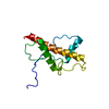

 Sample preparation
 Processing