- PDB-1u07: Crystal Structure of the 92-residue C-term. part of TonB with sig... -
+
Open data
ID or keywords:
Loading...
-
Basic information
Entry
Database: PDB / ID: 1u07
Title
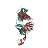
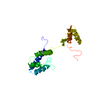
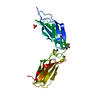
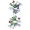
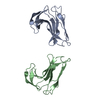
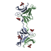
Crystal Structure of the 92-residue C-term. part of TonB with significant structural changes compared to shorter fragments
Components
TonB protein
Keywords
PROTEIN TRANSPORT / beta-hairpin
Function / homology
Function and homology information
receptor-mediated bacteriophage irreversible attachment to host cell / colicin transport / energy transducer activity / Metal ion assimilation from the host / cobalamin transport / intracellular monoatomic cation homeostasis / siderophore transport / Iron assimilation using enterobactin / plasma membrane protein complex / cell envelope ...receptor-mediated bacteriophage irreversible attachment to host cell / colicin transport / energy transducer activity / Metal ion assimilation from the host / cobalamin transport / intracellular monoatomic cation homeostasis / siderophore transport / Iron assimilation using enterobactin / plasma membrane protein complex / cell envelope / Heme assimilation / cell outer membrane / transmembrane transport / protein transport / outer membrane-bounded periplasmic space / intracellular iron ion homeostasis / protein domain specific binding / membrane / plasma membrane Similarity search - Function
Resolution: 1.08→1.1 Å / Redundancy: 1.73 % / Rmerge(I) obs: 0.519 / Mean I/σ(I) obs: 1.51 / Num. unique all: 2763 / % possible all: 78.5
-
Processing
Software
Name
Classification
MAR345
datacollection
XDS
datareduction
SOLVE
phasing
SHELXL-97
refinement
XDS
datascaling
Refinement
Method to determine structure: MAD / Resolution: 1.13→10 Å / Num. parameters: 15000 / Num. restraintsaints: 18590 / Isotropic thermal model: anisotropic / Cross valid method: free R / σ(F): -3 / σ(I): -3 StereochEM target val spec case: MOEWS & KRETSINGER, J.MOL.BIOL.91(1973)201-228 Stereochemistry target values: Engh & Huber Details: structure was solved with MAD on a Seleno-Met crystal at 2A. This entry is based on the high resolution data measured at 0.95372A wavelength. Anisotropic scaling applied by the method of ...Details: structure was solved with MAD on a Seleno-Met crystal at 2A. This entry is based on the high resolution data measured at 0.95372A wavelength. Anisotropic scaling applied by the method of parkin, MOEZZI & HOPE, J.APPL.CRYST.28(1995)53-56
Rfactor
Num. reflection
% reflection
Selection details
Rfree
0.183
2781
5 %
RANDOM
Rwork
0.137
-
-
-
all
0.139
55631
-
-
obs
0.139
55631
99.9 %
-
Refine analyze
Num. disordered residues: 15 / Occupancy sum hydrogen: 1445.46 / Occupancy sum non hydrogen: 1605.46
Refinement step
Cycle: LAST / Resolution: 1.13→10 Å
Protein
Nucleic acid
Ligand
Solvent
Total
Num. atoms
1464
0
0
198
1662
Refine LS restraints
Refine-ID
Type
Dev ideal
X-RAY DIFFRACTION
s_bond_d
0.014
X-RAY DIFFRACTION
s_angle_d
0.037
X-RAY DIFFRACTION
s_similar_dist
0
X-RAY DIFFRACTION
s_from_restr_planes
0.0269
X-RAY DIFFRACTION
s_zero_chiral_vol
0.077
X-RAY DIFFRACTION
s_non_zero_chiral_vol
0.101
X-RAY DIFFRACTION
s_anti_bump_dis_restr
0.027
X-RAY DIFFRACTION
s_rigid_bond_adp_cmpnt
0.005
X-RAY DIFFRACTION
s_similar_adp_cmpnt
0.066
X-RAY DIFFRACTION
s_approx_iso_adps
0.113
+
About Yorodumi
-
News
-
Feb 9, 2022. New format data for meta-information of EMDB entries
New format data for meta-information of EMDB entries
Version 3 of the EMDB header file is now the official format.
The previous official version 1.9 will be removed from the archive.
In the structure databanks used in Yorodumi, some data are registered as the other names, "COVID-19 virus" and "2019-nCoV". Here are the details of the virus and the list of structure data.
Jan 31, 2019. EMDB accession codes are about to change! (news from PDBe EMDB page)
EMDB accession codes are about to change! (news from PDBe EMDB page)
The allocation of 4 digits for EMDB accession codes will soon come to an end. Whilst these codes will remain in use, new EMDB accession codes will include an additional digit and will expand incrementally as the available range of codes is exhausted. The current 4-digit format prefixed with “EMD-” (i.e. EMD-XXXX) will advance to a 5-digit format (i.e. EMD-XXXXX), and so on. It is currently estimated that the 4-digit codes will be depleted around Spring 2019, at which point the 5-digit format will come into force.
The EM Navigator/Yorodumi systems omit the EMD- prefix.
Related info.:Q: What is EMD? / ID/Accession-code notation in Yorodumi/EM Navigator
Yorodumi is a browser for structure data from EMDB, PDB, SASBDB, etc.
This page is also the successor to EM Navigator detail page, and also detail information page/front-end page for Omokage search.
The word "yorodu" (or yorozu) is an old Japanese word meaning "ten thousand". "mi" (miru) is to see.
Related info.:EMDB / PDB / SASBDB / Comparison of 3 databanks / Yorodumi Search / Aug 31, 2016. New EM Navigator & Yorodumi / Yorodumi Papers / Jmol/JSmol / Function and homology information / Changes in new EM Navigator and Yorodumi
 Movie
Movie Controller

 Yorodumi
 Open data
 Basic information
 Components
 Keywords
 Function and homology information
 X-RAY DIFFRACTION /
  Authors
 Citation
 Structure visualization
 Downloads & links
 Other downloads
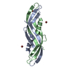


 PDBj
PDBj Assembly
 Mass: 18.015 Da / Num. of mol.: 198 / Source method: isolated from a natural source / Formula: H2O
 Sample preparation
 / Beamline: X06SA / Wavelength: 0.95372 Å
 Processing