 Movie
Movie Controller

[English] 日本語
 Yorodumi
- PDB-1tuw: Structural and Functional Analysis of Tetracenomycin F2 Cyclase f... -
+ Open data

- Basic information
| Entry | Database: PDB / ID: 1tuw | ||||||
|---|---|---|---|---|---|---|---|
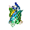
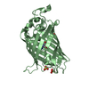
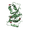
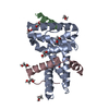
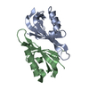
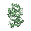
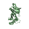
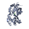
| Title | Structural and Functional Analysis of Tetracenomycin F2 Cyclase from Streptomyces glaucescens: A Type-II Polyketide Cyclase | ||||||
 Components Components | Tetracenomycin polyketide synthesis protein tcmI | ||||||
 Keywords Keywords | UNKNOWN FUNCTION / dimeric ??? ferredoxin-like fold tetracenomycin C biosynthesis | ||||||
| Function / homology |  Function and homology information Function and homology informationtetracenomycin F2 cyclase / polyketide biosynthetic process / antibiotic biosynthetic process / lyase activity Similarity search - Function | ||||||
| Biological species |  Streptomyces glaucescens (bacteria) Streptomyces glaucescens (bacteria) | ||||||
| Method |  X-RAY DIFFRACTION / MAD / Resolution: 1.9 Å X-RAY DIFFRACTION / MAD / Resolution: 1.9 Å | ||||||
 Authors Authors | Thompson, T.B. / Katayama, K. / Watanabe, K. / Hutchinson, C.R. / Rayment, I. | ||||||
 Citation Citation | Journal: J.Biol.Chem. / Year: 2004 Title: Structural and functional analysis of tetracenomycin F2 cyclase from Streptomyces glaucescens. A type II polyketide cyclase. Authors: Thompson, T.B. / Katayama, K. / Watanabe, K. / Hutchinson, C.R. / Rayment, I. | ||||||
| History |
|
- Structure visualization
| Structure viewer | Molecule: MolmilJmol/JSmol |
|---|
- Downloads & links
-Download
| PDBx/mmCIF format | 1tuw.cif.gz | 35.8 KB | Display | PDBx/mmCIF format |
|---|---|---|---|---|
| PDB format | pdb1tuw.ent.gz | 24.6 KB | Display | PDB format |
| PDBx/mmJSON format | 1tuw.json.gz | Tree view | PDBx/mmJSON format | |
| Others |  Other downloads Other downloads |
-Validation report
| Arichive directory | https://data.pdbj.org/pub/pdb/validation_reports/tu/1tuwftp://data.pdbj.org/pub/pdb/validation_reports/tu/1tuw | HTTPS FTP |
|---|
-Related structure data
| Similar structure data |
|---|

-Links
 PDBj
PDBj- Assembly
| Deposited unit | 
| ||||||||
|---|---|---|---|---|---|---|---|---|---|
| 1 | 
| ||||||||
| Unit cell |
|
-Components
| #1: Protein | Mass: 12877.448 Da / Num. of mol.: 1 Source method: isolated from a genetically manipulated source Source: (gene. exp.) Streptomyces glaucescens (bacteria) / Gene: TCMI / Species (production host): Escherichia coli / Production host: |
|---|---|
| #2: Chemical | ChemComp-SO4 /   Mass: 96.063 Da / Num. of mol.: 1 / Source method: obtained synthetically / Formula: SO4 Mass: 96.063 Da / Num. of mol.: 1 / Source method: obtained synthetically / Formula: SO4 |
| #3: Water | ChemComp-HOH /  Mass: 18.015 Da / Num. of mol.: 79 / Source method: isolated from a natural source / Formula: H2O Mass: 18.015 Da / Num. of mol.: 79 / Source method: isolated from a natural source / Formula: H2O |
-Experimental details
-Experiment
| Experiment | Method: X-RAY DIFFRACTION / Number of used crystals: 1 |
|---|
- Sample preparation
| Crystal | Density Matthews: 2.32 Å3/Da / Density % sol: 47 % |
|---|---|
| Crystal grow | Temperature: 278 K / Method: microbatch / pH: 7.5 Details: Crystals of tetracenomycin F2 cyclase were grown by microbatch from a solution composed of 2.2 M (NH4)2SO4, 3 mg/ml protein, 50 mM HEPES pH 7.5 at 4 C (11). Large crystals were obtained by ...Details: Crystals of tetracenomycin F2 cyclase were grown by microbatch from a solution composed of 2.2 M (NH4)2SO4, 3 mg/ml protein, 50 mM HEPES pH 7.5 at 4 C (11). Large crystals were obtained by macroseeding with small crystals (0.06 mm x 0.08 mm) washed in macroseeding 1.4 M (NH4)2SO4, 50 mM HEPES pH 7.5 to slightly dissolve the crystal surfaces. After 3 to 4 weeks the crystals attained a size of 0.3 x 0.3 x 0.6 mm. Crystals of the selenomethionine labeled protein were obtained in a similar manner., Micro batch, temperature 278K |
-Data collection
| Diffraction | Mean temperature: 273 K |
|---|---|
| Diffraction source | Source: ROTATING ANODE / Type: RIGAKU RU200 / Wavelength: 1.5418 |
| Detector | Type: SIEMENS HI-STAR / Detector: AREA DETECTOR / Date: Mar 1, 2000 / Details: gobel |
| Radiation | Monochromator: Gobel mirrors / Protocol: MAD / Monochromatic (M) / Laue (L): M / Scattering type: x-ray |
| Radiation wavelength | Wavelength: 1.5418 Å / Relative weight: 1 |
| Reflection | Resolution: 1.9→30 Å / Num. all: 11605 / Num. obs: 11355 / % possible obs: 92 % / Observed criterion σ(F): 0 / Observed criterion σ(I): 0 / Redundancy: 8.8 % / Rmerge(I) obs: 0.027 / Net I/σ(I): 23.1 |
| Reflection shell | Resolution: 1.9→2 Å / Rmerge(I) obs: 0.088 / Mean I/σ(I) obs: 5.4 / % possible all: 84 |
- Processing
| Software |
| ||||||||||||||||||||
|---|---|---|---|---|---|---|---|---|---|---|---|---|---|---|---|---|---|---|---|---|---|
| Refinement | Method to determine structure: MAD / Resolution: 1.9→30 Å / σ(F): 0 / σ(I): 0 / Stereochemistry target values: Engh & Huber
| ||||||||||||||||||||
| Refinement step | Cycle: LAST / Resolution: 1.9→30 Å
| ||||||||||||||||||||
| Refine LS restraints |
|
