 Movie
Movie Controller

[English] 日本語
 Yorodumi
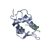
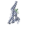
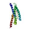
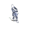
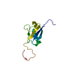
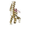
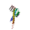
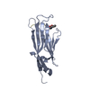
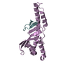
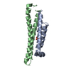
- PDB-1ttv: NMR Structure of a Complex Between MDM2 and a Small Molecule Inhibitor -
+ Open data

- Basic information
| Entry | Database: PDB / ID: 1ttv | ||||||
|---|---|---|---|---|---|---|---|
| Title | NMR Structure of a Complex Between MDM2 and a Small Molecule Inhibitor | ||||||
 Components Components | Ubiquitin-protein ligase E3 Mdm2 | ||||||
 Keywords Keywords | LIGASE / MDM2 / protein-protein interaction | ||||||
| Function / homology |  Function and homology information Function and homology informationpositive regulation of mitotic cell cycle / RING-type E3 ubiquitin transferase / p53 binding / ubiquitin protein ligase activity / regulation of gene expression / ubiquitin-dependent protein catabolic process / protein ubiquitination / apoptotic process / negative regulation of apoptotic process / nucleolus ...positive regulation of mitotic cell cycle / RING-type E3 ubiquitin transferase / p53 binding / ubiquitin protein ligase activity / regulation of gene expression / ubiquitin-dependent protein catabolic process / protein ubiquitination / apoptotic process / negative regulation of apoptotic process / nucleolus / zinc ion binding / nucleoplasm / identical protein binding / nucleus / cytoplasm Similarity search - Function | ||||||
| Biological species | |||||||
| Method | SOLUTION NMR / simulated annealing | ||||||
 Authors Authors | Fry, D.C. / Emerson, S.D. / Palme, S. / Vu, B.T. / Liu, C.M. / Podlaski, F. | ||||||
 Citation Citation | Journal: J.Biomol.Nmr / Year: 2004 Title: NMR structure of a complex between MDM2 and a small molecule inhibitor. Authors: Fry, D.C. / Emerson, S.D. / Palme, S. / Vu, B.T. / Liu, C.M. / Podlaski, F. | ||||||
| History |
| ||||||
| Remark 600 | HETEROGEN AUTHOR NOTED THAT THE piperizinyl moiety produced no NOEs, so the positions of its atoms ...HETEROGEN AUTHOR NOTED THAT THE piperizinyl moiety produced no NOEs, so the positions of its atoms could not be determined experimentally AND ARE NOT INCLUDED IN THE COORDINATES. |
- Structure visualization
| Structure viewer | Molecule: MolmilJmol/JSmol |
|---|
- Downloads & links
-Download
| PDBx/mmCIF format | 1ttv.cif.gz | 625.2 KB | Display | PDBx/mmCIF format |
|---|---|---|---|---|
| PDB format | pdb1ttv.ent.gz | 520 KB | Display | PDB format |
| PDBx/mmJSON format | 1ttv.json.gz | Tree view | PDBx/mmJSON format | |
| Others |  Other downloads Other downloads |
-Validation report
| Arichive directory | https://data.pdbj.org/pub/pdb/validation_reports/tt/1ttvftp://data.pdbj.org/pub/pdb/validation_reports/tt/1ttv | HTTPS FTP |
|---|
-Related structure data
| Related structure data | |
|---|---|
| Similar structure data |

-Links
 PDBj
PDBj

- Assembly
| Deposited unit | 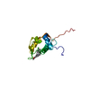
| |||||||||
|---|---|---|---|---|---|---|---|---|---|---|
| 1 |
| |||||||||
| NMR ensembles |
|
-Components
| #1: Protein | Mass: 12206.883 Da / Num. of mol.: 1 / Fragment: resiudes 13-119 / Mutation: I50L, P92H, L95I Source method: isolated from a genetically manipulated source Source: (gene. exp.)  References: UniProt: P56273, Ligases; Forming carbon-nitrogen bonds; Acid-amino-acid ligases (peptide synthases) |
|---|---|
| #2: Chemical | ChemComp-IMY / 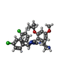  Mass: 567.506 Da / Num. of mol.: 1 / Source method: obtained synthetically / Formula: C30H32Cl2N4O3 Mass: 567.506 Da / Num. of mol.: 1 / Source method: obtained synthetically / Formula: C30H32Cl2N4O3 |
-Experimental details
-Experiment
| Experiment | Method: SOLUTION NMR | ||||||||||||||||
|---|---|---|---|---|---|---|---|---|---|---|---|---|---|---|---|---|---|
| NMR experiment |
|
- Sample preparation
| Details | Contents: 0.6mM 13C/15N-MDM2; 3.5mM inhibitor; 50mM d13-MES; 150mM KCl; 50mM d10-DTT; 1.5mM NaN3 Solvent system: 92% H2O, 8% D2O |
|---|---|
| Sample conditions | Ionic strength: 50mM d13-MES; 150mM KCl; 50mM d10-DTT; 1.5mM NaN3 pH: 7 / Pressure: ambient / Temperature: 293 K |
-NMR measurement
| Radiation | Protocol: SINGLE WAVELENGTH / Monochromatic (M) / Laue (L): M |
|---|---|
| Radiation wavelength | Relative weight: 1 |
| NMR spectrometer | Type: Varian INOVA / Manufacturer: Varian / Model: INOVA / Field strength: 600 MHz |
- Processing
| NMR software |
| ||||||||||||||||||||
|---|---|---|---|---|---|---|---|---|---|---|---|---|---|---|---|---|---|---|---|---|---|
| Refinement | Method: simulated annealing / Software ordinal: 1 Details: Structure calculations utilized 1169 NOE-based distance constraints, of which 54 were intermolecular; 176 dihedral angle constraints; and 48 hydrogen bond constraints. The piperazinyl moiety ...Details: Structure calculations utilized 1169 NOE-based distance constraints, of which 54 were intermolecular; 176 dihedral angle constraints; and 48 hydrogen bond constraints. The piperazinyl moiety of the inhibitor gave no NOEs, hence the positions of its atoms are not known and the stereochemistry is not rigorously established. | ||||||||||||||||||||
| NMR representative | Selection criteria: fewest violations | ||||||||||||||||||||
| NMR ensemble | Conformer selection criteria: structures with acceptable covalent geometry,structures with the least restraint violations Conformers calculated total number: 42 / Conformers submitted total number: 18 |
