 Movie
Movie Controller


+ Open data

- Basic information
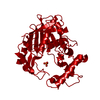
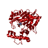
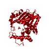
| Entry | Database: PDB / ID: 1tsl | ||||||
|---|---|---|---|---|---|---|---|
| Title | L. CASEI THYMIDYLATE SYNTHASE WITH SPECIES SPECIFIC INHIBITOR | ||||||
 Components Components | THYMIDYLATE SYNTHASE | ||||||
 Keywords Keywords | METHYLTRANSFERASE / SPECIES SPECIFICITY / STRUCTURE-BASED DRUG DESIGN / ANTIBIOTIC | ||||||
| Function / homology |  Function and homology information Function and homology informationthymidylate synthase / thymidylate synthase activity / dTTP biosynthetic process / dTMP biosynthetic process / methylation / cytosol Similarity search - Function | ||||||
| Biological species |  Lactobacillus casei (bacteria) Lactobacillus casei (bacteria) | ||||||
| Method |  X-RAY DIFFRACTION / DIFFERENCE FOURIER / Resolution: 2.5 Å X-RAY DIFFRACTION / DIFFERENCE FOURIER / Resolution: 2.5 Å | ||||||
 Authors Authors | Stout, T.J. / Stroud, R.M. | ||||||
 Citation Citation | Journal: Biochemistry / Year: 1999 Title: Structure-based design of inhibitors specific for bacterial thymidylate synthase. Authors: Stout, T.J. / Tondi, D. / Rinaldi, M. / Barlocco, D. / Pecorari, P. / Santi, D.V. / Kuntz, I.D. / Stroud, R.M. / Shoichet, B.K. / Costi, M.P. | ||||||
| History |
|
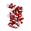
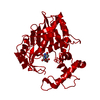
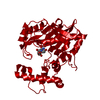
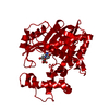
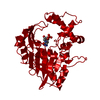
- Structure visualization
| Structure viewer | Molecule: MolmilJmol/JSmol |
|---|
- Downloads & links
-Download
| PDBx/mmCIF format | 1tsl.cif.gz | 85.2 KB | Display | PDBx/mmCIF format |
|---|---|---|---|---|
| PDB format | pdb1tsl.ent.gz | 64.4 KB | Display | PDB format |
| PDBx/mmJSON format | 1tsl.json.gz | Tree view | PDBx/mmJSON format | |
| Others |  Other downloads Other downloads |
-Validation report
| Arichive directory | https://data.pdbj.org/pub/pdb/validation_reports/ts/1tslftp://data.pdbj.org/pub/pdb/validation_reports/ts/1tsl | HTTPS FTP |
|---|
-Related structure data
| Related structure data | 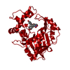 1tsmC  4tmsS S: Starting model for refinement C: citing same article ( |
|---|---|
| Similar structure data |

-Links
 PDBj
PDBj- Assembly
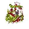
| Deposited unit | 
| ||||||||
|---|---|---|---|---|---|---|---|---|---|
| 1 | 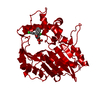
| ||||||||
| Unit cell |
|
-Components
| #1: Protein | Mass: 36630.453 Da / Num. of mol.: 1 Source method: isolated from a genetically manipulated source Source: (gene. exp.) Lactobacillus casei (bacteria) / Plasmid: PKPTSD / Production host: |
|---|---|
| #2: Chemical | ChemComp-PO4 /   Mass: 94.971 Da / Num. of mol.: 1 / Source method: obtained synthetically / Formula: PO4 Mass: 94.971 Da / Num. of mol.: 1 / Source method: obtained synthetically / Formula: PO4 |
| #3: Chemical | ChemComp-A15 / 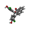  Mass: 437.272 Da / Num. of mol.: 1 / Source method: obtained synthetically / Formula: C24H14Cl2O4 Mass: 437.272 Da / Num. of mol.: 1 / Source method: obtained synthetically / Formula: C24H14Cl2O4 |
| #4: Water | ChemComp-HOH /  Mass: 18.015 Da / Num. of mol.: 313 / Source method: isolated from a natural source / Formula: H2O Mass: 18.015 Da / Num. of mol.: 313 / Source method: isolated from a natural source / Formula: H2O |
-Experimental details
-Experiment
| Experiment | Method: X-RAY DIFFRACTION / Number of used crystals: 1 |
|---|
- Sample preparation
| Crystal | Density Matthews: 2.94 Å3/Da / Density % sol: 58.11 % | ||||||||||||||||||||||||||||||||||||
|---|---|---|---|---|---|---|---|---|---|---|---|---|---|---|---|---|---|---|---|---|---|---|---|---|---|---|---|---|---|---|---|---|---|---|---|---|---|
| Crystal grow | Method: vapor diffusion, hanging drop / pH: 6.8 Details: 6 MG/ML PROTEIN, PH 7.4; 1% AMMONIUM SULFATE, 1 MM DTT, + 1 % SATURATED LIGAND-DMSO SOLUTION. HANGING DROP OVER 20 MM KPO4, PH 6.8 AND 1 MM DTT., vapor diffusion - hanging drop | ||||||||||||||||||||||||||||||||||||
| Crystal grow | *PLUS Method: vapor diffusion, hanging drop | ||||||||||||||||||||||||||||||||||||
| Components of the solutions | *PLUS
|
-Data collection
| Diffraction | Mean temperature: 287 K |
|---|---|
| Diffraction source | Source: ROTATING ANODE / Type: RIGAKU RUH2R / Wavelength: 1.5418 |
| Detector | Type: RIGAKU / Detector: IMAGE PLATE / Date: Mar 1, 1994 |
| Radiation | Monochromator: GRAPHITE(002) / Monochromatic (M) / Laue (L): M / Scattering type: x-ray |
| Radiation wavelength | Wavelength: 1.5418 Å / Relative weight: 1 |
| Reflection | Resolution: 2.5→48.6 Å / Num. obs: 9477 / % possible obs: 58.7 % / Observed criterion σ(I): 2 / Redundancy: 8.3 % / Biso Wilson estimate: 29.7 Å2 / Rmerge(I) obs: 0.0812 / Rsym value: 0.0812 / Net I/σ(I): 9.7 |
| Reflection shell | Resolution: 2.5→2.75 Å / Redundancy: 2.3 % / Rmerge(I) obs: 0.125 / Mean I/σ(I) obs: 3.5 / Rsym value: 0.125 / % possible all: 24.7 |
| Reflection | *PLUS Num. all: 29742 / Num. measured all: 78644 |
| Reflection shell | *PLUS % possible obs: 24.7 % |
- Processing
| Software |
| ||||||||||||||||||||||||||||||||||||||||||||||||||||||||||||||||||||||||||||||||
|---|---|---|---|---|---|---|---|---|---|---|---|---|---|---|---|---|---|---|---|---|---|---|---|---|---|---|---|---|---|---|---|---|---|---|---|---|---|---|---|---|---|---|---|---|---|---|---|---|---|---|---|---|---|---|---|---|---|---|---|---|---|---|---|---|---|---|---|---|---|---|---|---|---|---|---|---|---|---|---|---|---|
| Refinement | Method to determine structure: DIFFERENCE FOURIER Starting model: PDB ENTRY 4TMS Resolution: 2.5→8 Å / Rfactor Rfree error: 0.008 / Data cutoff high absF: 10000000 / Data cutoff low absF: 0.001 / Isotropic thermal model: RESTRAINED / Cross valid method: THROUGHOUT / σ(F): 2
| ||||||||||||||||||||||||||||||||||||||||||||||||||||||||||||||||||||||||||||||||
| Displacement parameters | Biso mean: 23 Å2 | ||||||||||||||||||||||||||||||||||||||||||||||||||||||||||||||||||||||||||||||||
| Refine analyze |
| ||||||||||||||||||||||||||||||||||||||||||||||||||||||||||||||||||||||||||||||||
| Refinement step | Cycle: LAST / Resolution: 2.5→8 Å
| ||||||||||||||||||||||||||||||||||||||||||||||||||||||||||||||||||||||||||||||||
| Refine LS restraints |
| ||||||||||||||||||||||||||||||||||||||||||||||||||||||||||||||||||||||||||||||||
| LS refinement shell | Resolution: 2.5→2.65 Å / Rfactor Rfree error: 0.051 / Total num. of bins used: 6
| ||||||||||||||||||||||||||||||||||||||||||||||||||||||||||||||||||||||||||||||||
| Xplor file |
| ||||||||||||||||||||||||||||||||||||||||||||||||||||||||||||||||||||||||||||||||
| Software | *PLUS Name: X-PLOR / Version: 3.1F / Classification: refinement | ||||||||||||||||||||||||||||||||||||||||||||||||||||||||||||||||||||||||||||||||
| Refinement | *PLUS | ||||||||||||||||||||||||||||||||||||||||||||||||||||||||||||||||||||||||||||||||
| Solvent computation | *PLUS | ||||||||||||||||||||||||||||||||||||||||||||||||||||||||||||||||||||||||||||||||
| Displacement parameters | *PLUS | ||||||||||||||||||||||||||||||||||||||||||||||||||||||||||||||||||||||||||||||||
| Refine LS restraints | *PLUS
| ||||||||||||||||||||||||||||||||||||||||||||||||||||||||||||||||||||||||||||||||
| LS refinement shell | *PLUS Rfactor obs: 0.222 |
