 Movie
Movie Controller

[English] 日本語
 Yorodumi
- PDB-1tpt: THREE-DIMENSIONAL STRUCTURE OF THYMIDINE PHOSPHORYLASE FROM ESCHE... -
+ Open data

- Basic information
| Entry | Database: PDB / ID: 1tpt | ||||||
|---|---|---|---|---|---|---|---|
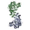
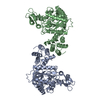
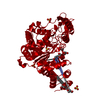
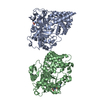
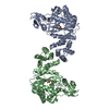
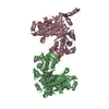
| Title | THREE-DIMENSIONAL STRUCTURE OF THYMIDINE PHOSPHORYLASE FROM ESCHERICHIA COLI AT 2.8 ANGSTROMS RESOLUTION | ||||||
 Components Components | THYMIDINE PHOSPHORYLASE | ||||||
 Keywords Keywords | THYMIDINE PHOSPHORYLASE | ||||||
| Function / homology |  Function and homology information Function and homology informationthymidine phosphorylase / pyrimidine nucleoside metabolic process / thymidine phosphorylase activity / thymidine metabolic process / pyrimidine nucleobase metabolic process / 1,4-alpha-oligoglucan phosphorylase activity / DNA damage response / membrane / cytosol Similarity search - Function | ||||||
| Biological species |  | ||||||
| Method |  X-RAY DIFFRACTION / Resolution: 2.8 Å X-RAY DIFFRACTION / Resolution: 2.8 Å | ||||||
 Authors Authors | Walter, M.R. / Cook, W.J. / Cole, L.B. / Short, S.A. / Koszalka, G.W. / Krenitsky, T.A. / Ealick, S.E. | ||||||
 Citation Citation | Journal: J.Biol.Chem. / Year: 1990 Title: Three-dimensional structure of thymidine phosphorylase from Escherichia coli at 2.8 A resolution. Authors: Walter, M.R. / Cook, W.J. / Cole, L.B. / Short, S.A. / Koszalka, G.W. / Krenitsky, T.A. / Ealick, S.E. | ||||||
| History |
|
- Structure visualization
| Structure viewer | Molecule: MolmilJmol/JSmol |
|---|
- Downloads & links
-Download
| PDBx/mmCIF format | 1tpt.cif.gz | 26 KB | Display | PDBx/mmCIF format |
|---|---|---|---|---|
| PDB format | pdb1tpt.ent.gz | 12.6 KB | Display | PDB format |
| PDBx/mmJSON format | 1tpt.json.gz | Tree view | PDBx/mmJSON format | |
| Others |  Other downloads Other downloads |
-Validation report
| Summary document | 1tpt_validation.pdf.gz | 313.5 KB | Display | wwPDB validaton report |
|---|---|---|---|---|
| Full document | 1tpt_full_validation.pdf.gz | 313.5 KB | Display | |
| Data in XML | 1tpt_validation.xml.gz | 1 KB | Display | |
| Data in CIF | 1tpt_validation.cif.gz | 4.9 KB | Display | |
| Arichive directory | https://data.pdbj.org/pub/pdb/validation_reports/tp/1tptftp://data.pdbj.org/pub/pdb/validation_reports/tp/1tpt | HTTPS FTP |
-Related structure data
| Similar structure data |
|---|
-Links
 PDBj
PDBj- Assembly
| Deposited unit | 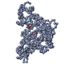
| ||||||||
|---|---|---|---|---|---|---|---|---|---|
| 1 | 
| ||||||||
| Unit cell |
|
-Components
| #1: Protein | Mass: 47240.988 Da / Num. of mol.: 1 Source method: isolated from a genetically manipulated source Source: (gene. exp.) |
|---|---|
| #2: Chemical | ChemComp-SO4 /   Mass: 96.063 Da / Num. of mol.: 1 / Source method: obtained synthetically / Formula: SO4 Mass: 96.063 Da / Num. of mol.: 1 / Source method: obtained synthetically / Formula: SO4 |
| #3: Chemical | ChemComp-TDR /   Mass: 126.113 Da / Num. of mol.: 1 / Source method: obtained synthetically / Formula: C5H6N2O2 Mass: 126.113 Da / Num. of mol.: 1 / Source method: obtained synthetically / Formula: C5H6N2O2 |
-Experimental details
-Experiment
| Experiment | Method: X-RAY DIFFRACTION |
|---|
- Sample preparation
| Crystal | Density Matthews: 3.16 Å3/Da / Density % sol: 61.06 % | ||||||||||||||||||||||||||||||
|---|---|---|---|---|---|---|---|---|---|---|---|---|---|---|---|---|---|---|---|---|---|---|---|---|---|---|---|---|---|---|---|
| Crystal grow | *PLUS pH: 5.5 / Method: vapor diffusion, hanging dropDetails: took from Cook, W.J.,(1987) J. Biol. Chem., 262, 3788. | ||||||||||||||||||||||||||||||
| Components of the solutions | *PLUS
|
-Data collection
| Reflection | *PLUS Highest resolution: 2.8 Å / Num. obs: 15034 / Num. measured all: 77819 / Rmerge F obs: 0.082 |
|---|
- Processing
| Software | Name: PROLSQ / Classification: refinement | ||||||||||||
|---|---|---|---|---|---|---|---|---|---|---|---|---|---|
| Refinement | Rfactor obs: 0.28 / Highest resolution: 2.8 Å | ||||||||||||
| Refinement step | Cycle: LAST / Highest resolution: 2.8 Å
| ||||||||||||
| Refine LS restraints |
| ||||||||||||
| Software | *PLUS Name: PROLSQ / Classification: refinement | ||||||||||||
| Refinement | *PLUS Rfactor obs: 0.28 | ||||||||||||
| Solvent computation | *PLUS | ||||||||||||
| Displacement parameters | *PLUS | ||||||||||||
| Refine LS restraints | *PLUS
|
