 Movie
Movie Controller

[English] 日本語
 Yorodumi
- PDB-1tb3: Crystal Structure Analysis of Recombinant Rat Kidney Long-chain H... -
+ Open data

- Basic information
| Entry | Database: PDB / ID: 1tb3 | ||||||
|---|---|---|---|---|---|---|---|
| Title | Crystal Structure Analysis of Recombinant Rat Kidney Long-chain Hydroxy Acid Oxidase | ||||||
 Components Components | Hydroxyacid oxidase 3 | ||||||
 Keywords Keywords | OXIDOREDUCTASE / long chain alpha-hydroxy acid oxidase / flavoprotein / oxidase | ||||||
| Function / homology |  Function and homology information Function and homology informationmandelate metabolic process / Peroxisomal lipid metabolism / Peroxisomal protein import / (S)-2-hydroxy-acid oxidase / (S)-2-hydroxy-acid oxidase activity / fatty acid oxidation / peroxisomal matrix / FMN binding / peroxisome / identical protein binding Similarity search - Function | ||||||
| Biological species |  | ||||||
| Method |  X-RAY DIFFRACTION / SYNCHROTRON / MOLECULAR REPLACEMENT / Resolution: 2.3 Å X-RAY DIFFRACTION / SYNCHROTRON / MOLECULAR REPLACEMENT / Resolution: 2.3 Å | ||||||
 Authors Authors | Cunane, L.M. / Barton, J.D. / Chen, Z.W. / Le, K.H.D. / Amar, D. / Lederer, F. / Mathews, F.S. | ||||||
 Citation Citation | Journal: Biochemistry / Year: 2005 Title: Crystal Structure Analysis of Recombinant Rat Kidney Long Chain Hydroxy Acid Oxidase. Authors: Cunane, L.M. / Barton, J.D. / Chen, Z.W. / Le, K.H.D. / Amar, D. / Lederer, F. / Mathews, F.S. #1: Journal: J.Mol.Biol. / Year: 1989Title: Refined structure of spinach glycolate oxidase at 2 A resolution. Authors: Lindqvist, Y. #2: Journal: J.Mol.Biol. / Year: 1990Title: Molecular structure of flavocytochrome b2 at 2.4 A resolution. Authors: Xia, Z. / Mathews, F.S. | ||||||
| History |
|
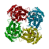
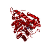
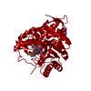
- Structure visualization
| Structure viewer | Molecule: MolmilJmol/JSmol |
|---|
- Downloads & links
-Download
| PDBx/mmCIF format | 1tb3.cif.gz | 534 KB | Display | PDBx/mmCIF format |
|---|---|---|---|---|
| PDB format | pdb1tb3.ent.gz | 437.3 KB | Display | PDB format |
| PDBx/mmJSON format | 1tb3.json.gz | Tree view | PDBx/mmJSON format | |
| Others |  Other downloads Other downloads |
-Validation report
| Arichive directory | https://data.pdbj.org/pub/pdb/validation_reports/tb/1tb3ftp://data.pdbj.org/pub/pdb/validation_reports/tb/1tb3 | HTTPS FTP |
|---|
-Related structure data
| Related structure data | 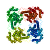 1goxS S: Starting model for refinement |
|---|---|
| Similar structure data |


-Links
 PDBj
PDBj


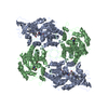
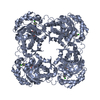
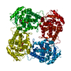
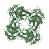
- Assembly
| Deposited unit | 
| ||||||||
|---|---|---|---|---|---|---|---|---|---|
| 1 | 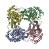
| ||||||||
| 2 | 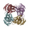
| ||||||||
| 3 |
| ||||||||
| Unit cell |
|
-Components
| #1: Protein | Mass: 39119.000 Da / Num. of mol.: 8 Source method: isolated from a genetically manipulated source Source: (gene. exp.)  #2: Chemical | ChemComp-FMN /   Mass: 456.344 Da / Num. of mol.: 8 / Source method: obtained synthetically / Formula: C17H21N4O9P Mass: 456.344 Da / Num. of mol.: 8 / Source method: obtained synthetically / Formula: C17H21N4O9P#3: Chemical | ChemComp-ACY /   Mass: 60.052 Da / Num. of mol.: 8 / Source method: obtained synthetically / Formula: C2H4O2 Mass: 60.052 Da / Num. of mol.: 8 / Source method: obtained synthetically / Formula: C2H4O2#4: Water | ChemComp-HOH / |  Mass: 18.015 Da / Num. of mol.: 900 / Source method: isolated from a natural source / Formula: H2O Mass: 18.015 Da / Num. of mol.: 900 / Source method: isolated from a natural source / Formula: H2O |
|---|
-Experimental details
-Experiment
| Experiment | Method: X-RAY DIFFRACTION / Number of used crystals: 1 |
|---|
- Sample preparation
| Crystal | Density Matthews: 3.1 Å3/Da / Density % sol: 60 % |
|---|---|
| Crystal grow | Temperature: 277 K / Method: vapor diffusion, sitting drop / pH: 6.5 Details: sodium acetate, sodium citrate, pH 6.5, VAPOR DIFFUSION, SITTING DROP, temperature 277K |
-Data collection
| Diffraction | Mean temperature: 100 K |
|---|---|
| Diffraction source | Source: SYNCHROTRON / Site: APS  / Beamline: 19-ID / Beamline: 19-ID |
| Detector | Type: SBC / Detector: CCD / Date: Apr 11, 1998 |
| Radiation | Protocol: SINGLE WAVELENGTH / Monochromatic (M) / Laue (L): M / Scattering type: x-ray |
| Radiation wavelength | Relative weight: 1 |
| Reflection | Resolution: 2.3→40 Å / Num. all: 171648 / Num. obs: 159633 / % possible obs: 93 % / Observed criterion σ(F): -3 / Observed criterion σ(I): -3 / Redundancy: 3.8 % / Biso Wilson estimate: 21.2 Å2 / Rmerge(I) obs: 0.05 / Net I/σ(I): 20.6 |
| Reflection shell | Resolution: 2.3→2.34 Å / Redundancy: 4.4 % / Rmerge(I) obs: 0.286 / Mean I/σ(I) obs: 3.4 / Num. unique all: 6770 / % possible all: 79.4 |
- Processing
| Software |
| |||||||||||||||||||||||||||||||||||||||||||||||||
|---|---|---|---|---|---|---|---|---|---|---|---|---|---|---|---|---|---|---|---|---|---|---|---|---|---|---|---|---|---|---|---|---|---|---|---|---|---|---|---|---|---|---|---|---|---|---|---|---|---|---|
| Refinement | Method to determine structure: MOLECULAR REPLACEMENT Starting model: PDB ENTRY 1GOX Resolution: 2.3→40 Å / Isotropic thermal model: Isotropic / Cross valid method: THROUGHOUT / σ(F): 0 / σ(I): 0 / Stereochemistry target values: Engh & Huber
| |||||||||||||||||||||||||||||||||||||||||||||||||
| Displacement parameters | Biso mean: 39.3 Å2 | |||||||||||||||||||||||||||||||||||||||||||||||||
| Refine analyze |
| |||||||||||||||||||||||||||||||||||||||||||||||||
| Refinement step | Cycle: LAST / Resolution: 2.3→40 Å
| |||||||||||||||||||||||||||||||||||||||||||||||||
| Refine LS restraints |
| |||||||||||||||||||||||||||||||||||||||||||||||||
| LS refinement shell |
|
