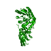
 ムービー
ムービー コントローラー


+ データを開く

- 基本情報
基本情報
| 登録情報 | データベース: PDB / ID: 1t8q | ||||||
|---|---|---|---|---|---|---|---|
| タイトル | Structural genomics, Crystal structure of Glycerophosphoryl diester phosphodiesterase from E. coli | ||||||
 要素 要素 | Glycerophosphoryl diester phosphodiesterase, periplasmic | ||||||
 キーワード キーワード | HYDROLASE / Phosphodiesterase / Structural genomics / PSI / Protein Structure Initiative / Midwest Center for Structural Genomics / MCSG | ||||||
| 機能・相同性 |  機能・相同性情報 機能・相同性情報glycerophosphodiester phosphodiesterase / glycerophospholipid catabolic process / glycerophosphodiester phosphodiesterase activity / glycerol metabolic process / periplasmic space / calcium ion binding / metal ion binding 類似検索 - 分子機能 | ||||||
| 生物種 |  | ||||||
| 手法 |  X線回折 / シンクロトロン / 多波長異常分散 / 解像度: 2 Å X線回折 / シンクロトロン / 多波長異常分散 / 解像度: 2 Å | ||||||
 データ登録者 データ登録者 | Zhang, R. / Kim, Y. / Dementieva, I. / Duke, N. / Stols, L. / Donnelly, M. / Joachimiak, A. / Midwest Center for Structural Genomics (MCSG) | ||||||
 引用 引用 | ジャーナル: To be Published タイトル: The crystal structure of Glycerophosphoryl diester phosphodiesterase from E. coli 著者: Zhang, R. / Kim, Y. / Dementieva, I. / Duke, N. / Stols, L. / Donnelly, M. / Joachimiak, A. | ||||||
| 履歴 |
|
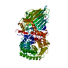
- 構造の表示
構造の表示
| 構造ビューア | 分子: MolmilJmol/JSmol |
|---|
- ダウンロードとリンク
-ダウンロード
| PDBx/mmCIF形式 | 1t8q.cif.gz | 303.1 KB | 表示 | PDBx/mmCIF形式 |
|---|---|---|---|---|
| PDB形式 | pdb1t8q.ent.gz | 242.8 KB | 表示 | PDB形式 |
| PDBx/mmJSON形式 | 1t8q.json.gz | ツリー表示 | PDBx/mmJSON形式 | |
| その他 |  その他のダウンロード その他のダウンロード |
-検証レポート
| アーカイブディレクトリ | https://data.pdbj.org/pub/pdb/validation_reports/t8/1t8qftp://data.pdbj.org/pub/pdb/validation_reports/t8/1t8q | HTTPS FTP |
|---|
-関連構造データ
| 類似構造データ | |
|---|---|
| その他のデータベース |


-リンク
 PDBj
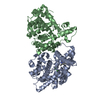
PDBj- 集合体
集合体
| 登録構造単位 | 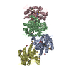
| ||||||||
|---|---|---|---|---|---|---|---|---|---|
| 1 | 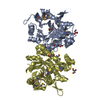
| ||||||||
| 2 | 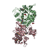
| ||||||||
| 単位格子 |
| ||||||||
| 詳細 | This protein existed as dimer. MolA & MolD, MolB & MolC represent two dimers in the asymmetric unit. |
-要素
| #1: タンパク質 | 分子量: 38944.211 Da / 分子数: 4 / 由来タイプ: 組換発現 / 由来: (組換発現) 参照: UniProt: P09394, glycerophosphodiester phosphodiesterase #2: 化合物 | ChemComp-MG /   分子量: 24.305 Da / 分子数: 4 / 由来タイプ: 合成 / 式: Mg 分子量: 24.305 Da / 分子数: 4 / 由来タイプ: 合成 / 式: Mg#3: 化合物 | ChemComp-GOL /   分子量: 92.094 Da / 分子数: 11 / 由来タイプ: 合成 / 式: C3H8O3 分子量: 92.094 Da / 分子数: 11 / 由来タイプ: 合成 / 式: C3H8O3#4: 水 | ChemComp-HOH / |  分子量: 18.015 Da / 分子数: 1160 / 由来タイプ: 天然 / 式: H2O 分子量: 18.015 Da / 分子数: 1160 / 由来タイプ: 天然 / 式: H2OHas protein modification | Y | |
|---|
-実験情報
-実験
| 実験 | 手法: X線回折 / 使用した結晶の数: 1 |
|---|
- 試料調製
試料調製
| 結晶 | マシュー密度: 2.958 Å3/Da / 溶媒含有率: 56.8 % / 解説: Friedel pairs were used |
|---|---|
| 結晶化 | 温度: 298 K / 手法: 蒸気拡散法, ハンギングドロップ法 / pH: 5.6 詳細: 16% PEG8000, 20% glycerol, 80 nM Na Cacodylate buffer, 0.16M Mg Acetate, pH 5.6, VAPOR DIFFUSION, HANGING DROP, temperature 298K |
-データ収集
| 回折 | 平均測定温度: 100 K | ||||||||||||
|---|---|---|---|---|---|---|---|---|---|---|---|---|---|
| 放射光源 | 由来: シンクロトロン / サイト: APS  / ビームライン: 19-ID / 波長: 0.9795, 0.9798, 0.94656 / ビームライン: 19-ID / 波長: 0.9795, 0.9798, 0.94656 | ||||||||||||
| 検出器 | タイプ: SBC-2 / 検出器: CCD / 日付: 2003年11月19日 / 詳細: mirrors | ||||||||||||
| 放射 | モノクロメーター: Si 111 channel / プロトコル: MAD / 単色(M)・ラウエ(L): M / 散乱光タイプ: x-ray | ||||||||||||
| 放射波長 |
| ||||||||||||
| 反射 | 解像度: 2→50 Å / Num. all: 239258 / Num. obs: 226817 / % possible obs: 94.8 % / Observed criterion σ(F): 2 / Observed criterion σ(I): 4 / 冗長度: 5.2 % / Biso Wilson estimate: 15.4 Å2 / Rmerge(I) obs: 0.086 / Net I/σ(I): 18.83 | ||||||||||||
| 反射 シェル | 解像度: 2→2.07 Å / 冗長度: 4.1 % / Rmerge(I) obs: 0.524 / Mean I/σ(I) obs: 1.68 / Num. unique all: 23943 / % possible all: 63.5 |
- 解析
解析
| ソフトウェア |
| ||||||||||||||||||||||||||||||||||||
|---|---|---|---|---|---|---|---|---|---|---|---|---|---|---|---|---|---|---|---|---|---|---|---|---|---|---|---|---|---|---|---|---|---|---|---|---|---|
| 精密化 | 構造決定の手法: 多波長異常分散 / 解像度: 2→43.56 Å / Rfactor Rfree error: 0.003 / Data cutoff high absF: 190352.26 / Data cutoff low absF: 0 / Isotropic thermal model: RESTRAINED / 交差検証法: THROUGHOUT / σ(F): 0 / σ(I): 0 / 立体化学のターゲット値: Engh & Huber
| ||||||||||||||||||||||||||||||||||||
| 溶媒の処理 | 溶媒モデル: FLAT MODEL / Bsol: 44.5213 Å2 / ksol: 0.343282 e/Å3 | ||||||||||||||||||||||||||||||||||||
| 原子変位パラメータ | Biso mean: 35.3 Å2
| ||||||||||||||||||||||||||||||||||||
| Refine analyze |
| ||||||||||||||||||||||||||||||||||||
| 精密化ステップ | サイクル: LAST / 解像度: 2→43.56 Å
| ||||||||||||||||||||||||||||||||||||
| 拘束条件 |
| ||||||||||||||||||||||||||||||||||||
| LS精密化 シェル | 解像度: 2→2.13 Å / Rfactor Rfree error: 0.01 / Total num. of bins used: 6
| ||||||||||||||||||||||||||||||||||||
| Xplor file |
|
