Resolution: 1.75→1.88 Å / Redundancy: 3.1 % / Rmerge(I) obs: 0.096 / Mean I/σ(I) obs: 9.2 / Num. unique all: 3509 / % possible all: 64.4
-
Processing
Software
Name
Version
Classification
XDS
datascaling
XDS
datareduction
SHARP
phasing
CNS
1.1
refinement
Refinement
Method to determine structure: SIRAS / Resolution: 1.8→25 Å / Isotropic thermal model: Isotropic / Cross valid method: THROUGHOUT / σ(F): 0 / σ(I): 0 / Stereochemistry target values: Engh & Huber Details: Cristallographic conjugate gradient refinement using maximum likelihood target for amplitudes. The residue Asp 124 has unusual Phi and Psi angles. The author maintains that this conformation ...Details: Cristallographic conjugate gradient refinement using maximum likelihood target for amplitudes. The residue Asp 124 has unusual Phi and Psi angles. The author maintains that this conformation is important for the function of the protein
Rfactor
Num. reflection
% reflection
Selection details
Rfree
0.21
2207
8.3 %
Random
Rwork
0.18
-
-
-
all
-
22062
-
-
obs
-
22062
83.1 %
-
Displacement parameters
Biso mean: 25.76 Å2
Baniso -1
Baniso -2
Baniso -3
1-
-10.105 Å2
0 Å2
0 Å2
2-
-
2.025 Å2
0 Å2
3-
-
-
8.08 Å2
Refine analyze
Free
Obs
Luzzati coordinate error
0.23 Å
0.2 Å
Luzzati d res low
-
5 Å
Luzzati sigma a
0.19 Å
0.16 Å
Refinement step
Cycle: LAST / Resolution: 1.8→25 Å
Protein
Nucleic acid
Ligand
Solvent
Total
Num. atoms
2096
0
9
250
2355
Refine LS restraints
Refine-ID
Type
Dev ideal
X-RAY DIFFRACTION
c_bond_d
0.009
X-RAY DIFFRACTION
c_angle_deg
1.55
X-RAY DIFFRACTION
c_dihedral_angle_d
25.5
X-RAY DIFFRACTION
c_improper_angle_d
0.927
X-RAY DIFFRACTION
c_mcbond_it
1.728
X-RAY DIFFRACTION
c_mcangle_it
2.485
X-RAY DIFFRACTION
c_scbond_it
2.653
X-RAY DIFFRACTION
c_scangle_it
3.675
LS refinement shell
Resolution: 1.8→1.86 Å / Total num. of bins used: 10
Rfactor
Num. reflection
% reflection
Rfree
0.2895
176
0.0684 %
Rwork
0.2639
1578
-
obs
-
1754
0.6777 %
+
About Yorodumi
-
News
-
Feb 9, 2022. New format data for meta-information of EMDB entries
New format data for meta-information of EMDB entries
Version 3 of the EMDB header file is now the official format.
The previous official version 1.9 will be removed from the archive.
In the structure databanks used in Yorodumi, some data are registered as the other names, "COVID-19 virus" and "2019-nCoV". Here are the details of the virus and the list of structure data.
Jan 31, 2019. EMDB accession codes are about to change! (news from PDBe EMDB page)
EMDB accession codes are about to change! (news from PDBe EMDB page)
The allocation of 4 digits for EMDB accession codes will soon come to an end. Whilst these codes will remain in use, new EMDB accession codes will include an additional digit and will expand incrementally as the available range of codes is exhausted. The current 4-digit format prefixed with “EMD-” (i.e. EMD-XXXX) will advance to a 5-digit format (i.e. EMD-XXXXX), and so on. It is currently estimated that the 4-digit codes will be depleted around Spring 2019, at which point the 5-digit format will come into force.
The EM Navigator/Yorodumi systems omit the EMD- prefix.
Related info.:Q: What is EMD? / ID/Accession-code notation in Yorodumi/EM Navigator
Yorodumi is a browser for structure data from EMDB, PDB, SASBDB, etc.
This page is also the successor to EM Navigator detail page, and also detail information page/front-end page for Omokage search.
The word "yorodu" (or yorozu) is an old Japanese word meaning "ten thousand". "mi" (miru) is to see.
Related info.:EMDB / PDB / SASBDB / Comparison of 3 databanks / Yorodumi Search / Aug 31, 2016. New EM Navigator & Yorodumi / Yorodumi Papers / Jmol/JSmol / Function and homology information / Changes in new EM Navigator and Yorodumi
 Movie
Movie Controller

 Yorodumi
 Open data
 Basic information
 Components
 Keywords
 Function and homology information
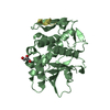
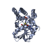
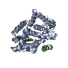
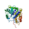
 Geobacillus stearothermophilus (bacteria)
 X-RAY DIFFRACTION /
  Authors
 Citation
 Structure visualization
 Downloads & links
 Other downloads
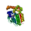


 PDBj
PDBj


 Assembly
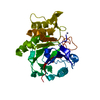

 Mass: 65.409 Da / Num. of mol.: 1 / Source method: obtained synthetically / Formula: Zn
 Mass: 78.133 Da / Num. of mol.: 2 / Source method: obtained synthetically / Formula: C2H6OS
 Mass: 18.015 Da / Num. of mol.: 239 / Source method: isolated from a natural source / Formula: H2O
 Sample preparation
 / Beamline: 5ID-B / Wavelength: 0.97985 Å
 Processing