 Movie
Movie Controller

[English] 日本語
 Yorodumi
- PDB-1t39: HUMAN O6-ALKYLGUANINE-DNA ALKYLTRANSFERASE COVALENTLY CROSSLINKED... -
+ Open data

- Basic information
| Entry | Database: PDB / ID: 1t39 | ||||||
|---|---|---|---|---|---|---|---|
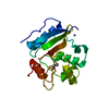
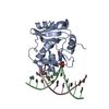
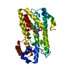
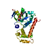
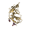
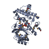
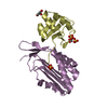
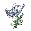
| Title | HUMAN O6-ALKYLGUANINE-DNA ALKYLTRANSFERASE COVALENTLY CROSSLINKED TO DNA | ||||||
 Components Components |
| ||||||
 Keywords Keywords | TRANSFERASE/DNA / ALKYLTRANSFERASE / METHYLTRANSFERASE / DNA REPAIR / HELIX-TURN-HELIX / TRANSFERASE-DNA COMPLEX | ||||||
| Function / homology |  Function and homology information Function and homology informationMGMT-mediated DNA damage reversal / methylated-DNA-[protein]-cysteine S-methyltransferase / methylated-DNA-[protein]-cysteine S-methyltransferase activity / DNA-methyltransferase activity / positive regulation of double-strand break repair / methyltransferase activity / methylation / DNA repair / negative regulation of apoptotic process / DNA binding ...MGMT-mediated DNA damage reversal / methylated-DNA-[protein]-cysteine S-methyltransferase / methylated-DNA-[protein]-cysteine S-methyltransferase activity / DNA-methyltransferase activity / positive regulation of double-strand break repair / methyltransferase activity / methylation / DNA repair / negative regulation of apoptotic process / DNA binding / nucleoplasm / membrane / metal ion binding / nucleus Similarity search - Function | ||||||
| Biological species |  Homo sapiens (human) Homo sapiens (human) | ||||||
| Method |  X-RAY DIFFRACTION / SYNCHROTRON / MOLECULAR REPLACEMENT / Resolution: 3.3 Å X-RAY DIFFRACTION / SYNCHROTRON / MOLECULAR REPLACEMENT / Resolution: 3.3 Å | ||||||
 Authors Authors | Daniels, D.S. / Woo, T.T. / Luu, K.X. / Noll, D.M. / Clarke, N.D. / Pegg, A.E. / Tainer, J.A. | ||||||
 Citation Citation | Journal: Nat.Struct.Mol.Biol. / Year: 2004 Title: DNA binding and nucleotide flipping by the human DNA repair protein AGT. Authors: Daniels, D.S. / Woo, T.T. / Luu, K.X. / Noll, D.M. / Clarke, N.D. / Pegg, A.E. / Tainer, J.A. #1: Journal: Embo J. / Year: 2000Title: Active and alkylated human AGT structures: a novel zinc site, inhibitor and extrahelical base binding Authors: Daniels, D.S. / Mol, C.D. / Arvai, A.S. / Kanugula, S. / Pegg, A.E. / Tainer, J.A. #2: Journal: Mutat.Res. / Year: 2000Title: Conserved structural motifs governing the stoichiometric repair of alkylated DNA by O(6)-alkylguanine-DNA alkyltransferase Authors: Daniels, D.S. / Tainer, J.A. | ||||||
| History |
| ||||||
| Remark 999 | SEQUENCE CYS 145 residues are COVALENTly CHEMICAL CROSSLINK TO E1X 7 residues |
- Structure visualization
| Structure viewer | Molecule: MolmilJmol/JSmol |
|---|
- Downloads & links
-Download
| PDBx/mmCIF format | 1t39.cif.gz | 92.6 KB | Display | PDBx/mmCIF format |
|---|---|---|---|---|
| PDB format | pdb1t39.ent.gz | 67.4 KB | Display | PDB format |
| PDBx/mmJSON format | 1t39.json.gz | Tree view | PDBx/mmJSON format | |
| Others |  Other downloads Other downloads |
-Validation report
| Arichive directory | https://data.pdbj.org/pub/pdb/validation_reports/t3/1t39ftp://data.pdbj.org/pub/pdb/validation_reports/t3/1t39 | HTTPS FTP |
|---|
-Related structure data
| Related structure data |  1t38C  1eh6S S: Starting model for refinement C: citing same article ( |
|---|---|
| Similar structure data |


-Links
 PDBj
PDBj


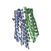
- Assembly
| Deposited unit | 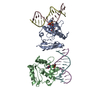
| ||||||||
|---|---|---|---|---|---|---|---|---|---|
| 1 | 
| ||||||||
| 2 | 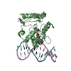
| ||||||||
| Unit cell |
|
-Components
| #1: DNA chain | Mass: 4020.648 Da / Num. of mol.: 2 / Source method: obtained synthetically #2: DNA chain | Mass: 3951.586 Da / Num. of mol.: 2 / Source method: obtained synthetically #3: Protein | Mass: 20355.473 Da / Num. of mol.: 2 Source method: isolated from a genetically manipulated source Source: (gene. exp.) Homo sapiens (human) / Gene: MGMT / Plasmid: pQE30 / Production host:  References: UniProt: P16455, methylated-DNA-[protein]-cysteine S-methyltransferase Has protein modification | Y | |
|---|
-Experimental details
-Experiment
| Experiment | Method: X-RAY DIFFRACTION / Number of used crystals: 1 |
|---|
- Sample preparation
| Crystal | Density Matthews: 2.86 Å3/Da / Density % sol: 57.1 % | ||||||||||||||||||||||||||||||||
|---|---|---|---|---|---|---|---|---|---|---|---|---|---|---|---|---|---|---|---|---|---|---|---|---|---|---|---|---|---|---|---|---|---|
| Crystal grow | Temperature: 298 K / Method: vapor diffusion, hanging drop / pH: 6 Details: PEG 8000, MES, calcium acetate, pH 6.0, VAPOR DIFFUSION, HANGING DROP, temperature 298K | ||||||||||||||||||||||||||||||||
| Components of the solutions |
|
-Data collection
| Diffraction | Mean temperature: 200 K |
|---|---|
| Diffraction source | Source: SYNCHROTRON / Site: SSRL  / Beamline: BL9-1 / Wavelength: 0.98 Å / Beamline: BL9-1 / Wavelength: 0.98 Å |
| Detector | Date: Jun 28, 2002 |
| Radiation | Protocol: SINGLE WAVELENGTH / Monochromatic (M) / Laue (L): M / Scattering type: x-ray |
| Radiation wavelength | Wavelength: 0.98 Å / Relative weight: 1 |
| Reflection | Resolution: 3.3→53 Å / Num. obs: 9362 |
| Reflection shell | Highest resolution: 3.3 Å / Num. unique all: 9362 |
- Processing
| Software |
| ||||||||||||||||||||||||||||||||||||||||||||||||||||||||||||
|---|---|---|---|---|---|---|---|---|---|---|---|---|---|---|---|---|---|---|---|---|---|---|---|---|---|---|---|---|---|---|---|---|---|---|---|---|---|---|---|---|---|---|---|---|---|---|---|---|---|---|---|---|---|---|---|---|---|---|---|---|---|
| Refinement | Method to determine structure: MOLECULAR REPLACEMENT Starting model: PDB ENTRY 1EH6 Resolution: 3.3→27.77 Å / Rfactor Rfree error: 0.011 / Data cutoff high absF: 8413320.97 / Data cutoff high rms absF: 8413320.97 / Data cutoff low absF: 0 / Isotropic thermal model: GROUP / Cross valid method: THROUGHOUT / σ(F): 0 / Stereochemistry target values: Engh & Huber
| ||||||||||||||||||||||||||||||||||||||||||||||||||||||||||||
| Solvent computation | Solvent model: FLAT MODEL / Bsol: 50.4823 Å2 / ksol: 0.185184 e/Å3 | ||||||||||||||||||||||||||||||||||||||||||||||||||||||||||||
| Refine analyze |
| ||||||||||||||||||||||||||||||||||||||||||||||||||||||||||||
| Refinement step | Cycle: LAST / Resolution: 3.3→27.77 Å
| ||||||||||||||||||||||||||||||||||||||||||||||||||||||||||||
| Refine LS restraints |
| ||||||||||||||||||||||||||||||||||||||||||||||||||||||||||||
| LS refinement shell | Highest resolution: 3.3 Å / Total num. of bins used: 6 /
| ||||||||||||||||||||||||||||||||||||||||||||||||||||||||||||
| Xplor file |
|
