[English] 日本語
 Yorodumi
- PDB-1sx0: Solution NMR Structure and X-Ray Absorption Analysis of the C-Ter... -
+ Open data
- Basic information
| Entry | Database: PDB / ID: 1sx0 | ||||||
|---|---|---|---|---|---|---|---|
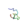
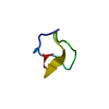
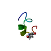
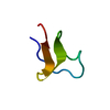
| Title | Solution NMR Structure and X-Ray Absorption Analysis of the C-Terminal Zinc-Binding Domain of the SecA ATPase | ||||||
 Components Components | SecA | ||||||
 Keywords Keywords | PROTEIN TRANSPORT / zinc / metal ion / tetrahedral coordination / no secondary structure / structural zinc coordination | ||||||
| Method | SOLUTION NMR / simulated annealing | ||||||
 Authors Authors | Dempsey, B.R. / Wrona, M. / Moulin, J.M. / Gloor, G.B. / Jalilehvand, F. / Lajoie, G. / Shaw, G.S. / Shilton, B.H. | ||||||
 Citation Citation |  Journal: Biochemistry / Year: 2004 Journal: Biochemistry / Year: 2004Title: Solution NMR Structure and X-ray Absorption Analysis of the C-Terminal Zinc-Binding Domain of the SecA ATPase. Authors: Dempsey, B.R. / Wrona, M. / Moulin, J.M. / Gloor, G.B. / Jalilehvand, F. / Lajoie, G. / Shaw, G.S. / Shilton, B.H. | ||||||
| History |
|
- Structure visualization
| Structure viewer | Molecule:  Molmil Molmil Jmol/JSmol Jmol/JSmol |
|---|
- Downloads & links
- Download
| PDBx/mmCIF format |  1sx0.cif.gz 1sx0.cif.gz | 117.4 KB | Display |  PDBx/mmCIF format PDBx/mmCIF format |
|---|---|---|---|---|
| PDB format |  pdb1sx0.ent.gz pdb1sx0.ent.gz | 82.3 KB | Display |  PDB format PDB format |
| PDBx/mmJSON format |  1sx0.json.gz 1sx0.json.gz | Tree view |  PDBx/mmJSON format PDBx/mmJSON format | |
| Others |  Other downloads Other downloads |
-Validation report
| Arichive directory |  https://data.pdbj.org/pub/pdb/validation_reports/sx/1sx0 https://data.pdbj.org/pub/pdb/validation_reports/sx/1sx0 ftp://data.pdbj.org/pub/pdb/validation_reports/sx/1sx0 ftp://data.pdbj.org/pub/pdb/validation_reports/sx/1sx0 | HTTPS FTP |
|---|
-Related structure data
- Links
Links
- Assembly
| Deposited unit | 
| |||||||||
|---|---|---|---|---|---|---|---|---|---|---|
| 1 |
| |||||||||
| NMR ensembles |
|
- Components
| #1: Protein/peptide | Mass: 2425.836 Da / Num. of mol.: 1 / Fragment: C-terminal Zinc Binding Domain / Source method: obtained synthetically Details: Solid phase peptide synthesis, N-terminally acetylated. The sequence of this peptide naturally exists in Escherichia coli |
|---|
-Experimental details
-Experiment
| Experiment | Method: SOLUTION NMR | ||||||||||||||||
|---|---|---|---|---|---|---|---|---|---|---|---|---|---|---|---|---|---|
| NMR experiment |
| ||||||||||||||||
| NMR details | Text: This structure was determined using standard two-dimensional 1H NMR techniques. This set of structures is the calculation of the initial fold of the domain without using restraints for zinc ...Text: This structure was determined using standard two-dimensional 1H NMR techniques. This set of structures is the calculation of the initial fold of the domain without using restraints for zinc coordination. A second set of structures has been deposited that is a refinement of this fold using zinc coordination restraints based on EXAFS data for this domain. |
- Sample preparation
| Details |
| |||||||||||||||
|---|---|---|---|---|---|---|---|---|---|---|---|---|---|---|---|---|
| Sample conditions |
|
-NMR measurement
| Radiation | Protocol: SINGLE WAVELENGTH / Monochromatic (M) / Laue (L): M | |||||||||||||||
|---|---|---|---|---|---|---|---|---|---|---|---|---|---|---|---|---|
| Radiation wavelength | Relative weight: 1 | |||||||||||||||
| NMR spectrometer |
|
- Processing
| NMR software |
| ||||||||||||||||||||||||
|---|---|---|---|---|---|---|---|---|---|---|---|---|---|---|---|---|---|---|---|---|---|---|---|---|---|
| Refinement | Method: simulated annealing / Software ordinal: 1 Details: structures based on 307 restraints, 274 are NOE-derived distance restraints, 33 are dihedral angle restraints. | ||||||||||||||||||||||||
| NMR representative | Selection criteria: closest to the average | ||||||||||||||||||||||||
| NMR ensemble | Conformer selection criteria: structures with the least restraint violations Conformers calculated total number: 100 / Conformers submitted total number: 20 |
 Movie
Movie Controller


 PDBj
PDBj NMRPipe