 Movie
Movie Controller

[English] 日本語
 Yorodumi
- PDB-1stb: ACCOMMODATION OF INSERTION MUTATIONS ON THE SURFACE AND IN THE IN... -
+ Open data

- Basic information
| Entry | Database: PDB / ID: 1stb | ||||||
|---|---|---|---|---|---|---|---|
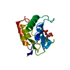
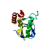
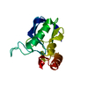
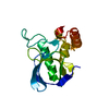
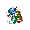
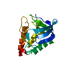
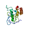
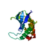
| Title | ACCOMMODATION OF INSERTION MUTATIONS ON THE SURFACE AND IN THE INTERIOR OF STAPHYLOCOCCAL NUCLEASE | ||||||
 Components Components | STAPHYLOCOCCAL NUCLEASE | ||||||
 Keywords Keywords | HYDROLASE(PHOSPHORIC DIESTER) | ||||||
| Function / homology |  Function and homology information Function and homology informationmicrococcal nuclease / : / nucleic acid binding / extracellular region / metal ion binding / membrane Similarity search - Function | ||||||
| Biological species |   Staphylococcus aureus (bacteria) Staphylococcus aureus (bacteria) | ||||||
| Method |  X-RAY DIFFRACTION / Resolution: 2 Å X-RAY DIFFRACTION / Resolution: 2 Å | ||||||
 Authors Authors | Quirk, S. / Gittis, A. / Keefe, L.J. / Lattman, E.E. | ||||||
 Citation Citation | Journal: Protein Sci. / Year: 1994 Title: Accommodation of insertion mutations on the surface and in the interior of staphylococcal nuclease. Authors: Keefe, L.J. / Quirk, S. / Gittis, A. / Sondek, J. / Lattman, E.E. #1: Journal: Proc.Natl.Acad.Sci.USA / Year: 1993Title: The Alpha Aneurism: A Structural Motif Revealed in an Insertion Mutant of Staphylococcal Nuclease Authors: Keefe, L.J. / Sondek, J. / Shortle, D. / Lattman, E.E. | ||||||
| History |
|
- Structure visualization
| Structure viewer | Molecule: MolmilJmol/JSmol |
|---|
- Downloads & links
-Download
| PDBx/mmCIF format | 1stb.cif.gz | 42.4 KB | Display | PDBx/mmCIF format |
|---|---|---|---|---|
| PDB format | pdb1stb.ent.gz | 28.5 KB | Display | PDB format |
| PDBx/mmJSON format | 1stb.json.gz | Tree view | PDBx/mmJSON format | |
| Others |  Other downloads Other downloads |
-Validation report
| Arichive directory | https://data.pdbj.org/pub/pdb/validation_reports/st/1stbftp://data.pdbj.org/pub/pdb/validation_reports/st/1stb | HTTPS FTP |
|---|
-Related structure data


-Links
 PDBj
PDBj- Assembly
| Deposited unit | 
| ||||||||
|---|---|---|---|---|---|---|---|---|---|
| 1 |
| ||||||||
| Unit cell |
| ||||||||
| Atom site foot note | 1: CIS PROLINE - PRO 117 |
-Components
| #1: Protein | Mass: 16956.486 Da / Num. of mol.: 1 Source method: isolated from a genetically manipulated source Source: (gene. exp.) Staphylococcus aureus (bacteria) / References: UniProt: P00644, micrococcal nuclease |
|---|---|
| #2: Chemical | ChemComp-CA /   Mass: 40.078 Da / Num. of mol.: 1 / Source method: obtained synthetically / Formula: Ca Mass: 40.078 Da / Num. of mol.: 1 / Source method: obtained synthetically / Formula: Ca |
| #3: Chemical | ChemComp-THP /   Type: DNA linking / Mass: 402.188 Da / Num. of mol.: 1 / Source method: obtained synthetically / Formula: C10H16N2O11P2 Type: DNA linking / Mass: 402.188 Da / Num. of mol.: 1 / Source method: obtained synthetically / Formula: C10H16N2O11P2 |
| #4: Water | ChemComp-HOH /  Mass: 18.015 Da / Num. of mol.: 39 / Source method: isolated from a natural source / Formula: H2O Mass: 18.015 Da / Num. of mol.: 39 / Source method: isolated from a natural source / Formula: H2O |
| Compound details | THE INSERTED LEUCINE RESIDUE IS ACCOMMODATED IN A REGISTER SHIFT THAT CHANGES THE CONFORMATION OF ...THE INSERTED LEUCINE RESIDUE IS ACCOMMODAT |
-Experimental details
-Experiment
| Experiment | Method: X-RAY DIFFRACTION |
|---|
- Sample preparation
| Crystal | Density Matthews: 2.07 Å3/Da / Density % sol: 40.58 % | ||||||||||||||||||||||||||||||||||||||||||||||||||||||
|---|---|---|---|---|---|---|---|---|---|---|---|---|---|---|---|---|---|---|---|---|---|---|---|---|---|---|---|---|---|---|---|---|---|---|---|---|---|---|---|---|---|---|---|---|---|---|---|---|---|---|---|---|---|---|---|
| Crystal grow | *PLUS Temperature: 4 ℃ / pH: 8.15 / Method: vapor diffusionDetails: taken from Loll, P.J. and Lattman, E.E. (1989). Proteins Struct. Funct. Genet., 5, 183-201. | ||||||||||||||||||||||||||||||||||||||||||||||||||||||
| Components of the solutions | *PLUS
|
-Data collection
| Reflection | *PLUS Highest resolution: 2 Å / Num. obs: 7854 / % possible obs: 85.1 % / Num. measured all: 42548 / Rmerge(I) obs: 0.042 |
|---|
- Processing
| Software |
| ||||||||||||||||||||||||||||||||||||||||||||||||||||||||||||
|---|---|---|---|---|---|---|---|---|---|---|---|---|---|---|---|---|---|---|---|---|---|---|---|---|---|---|---|---|---|---|---|---|---|---|---|---|---|---|---|---|---|---|---|---|---|---|---|---|---|---|---|---|---|---|---|---|---|---|---|---|---|
| Refinement | Resolution: 2→6 Å / σ(F): 4 /
| ||||||||||||||||||||||||||||||||||||||||||||||||||||||||||||
| Refinement step | Cycle: LAST / Resolution: 2→6 Å
| ||||||||||||||||||||||||||||||||||||||||||||||||||||||||||||
| Refine LS restraints |
| ||||||||||||||||||||||||||||||||||||||||||||||||||||||||||||
| Refinement | *PLUS Rfactor obs: 0.196 / Rfactor Rwork: 0.196 | ||||||||||||||||||||||||||||||||||||||||||||||||||||||||||||
| Solvent computation | *PLUS | ||||||||||||||||||||||||||||||||||||||||||||||||||||||||||||
| Displacement parameters | *PLUS | ||||||||||||||||||||||||||||||||||||||||||||||||||||||||||||
| Refine LS restraints | *PLUS
|
