 Movie
Movie Controller


+ Open data

- Basic information
| Entry | Database: PDB / ID: 1snb | ||||||
|---|---|---|---|---|---|---|---|
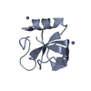
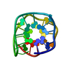
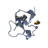
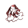
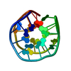
| Title | STRUCTURE OF SCORPION NEUROTOXIN BMK M8 | ||||||
 Components Components | NEUROTOXIN BMK M8 | ||||||
 Keywords Keywords | NEUROTOXIN / SODIUM CHANNEL INHIBITOR / SCORPION | ||||||
| Function / homology |  Function and homology information Function and homology informationsodium channel inhibitor activity / defense response / toxin activity / extracellular region Similarity search - Function | ||||||
| Biological species |  Mesobuthus martensii (Chinese scorpion) Mesobuthus martensii (Chinese scorpion) | ||||||
| Method |  X-RAY DIFFRACTION / MOLECULAR REPLACEMENT / Resolution: 1.9 Å X-RAY DIFFRACTION / MOLECULAR REPLACEMENT / Resolution: 1.9 Å | ||||||
 Authors Authors | Wang, D.C. / Zeng, Z.H. / Li, H.M. | ||||||
 Citation Citation | Journal: J.Mol.Biol. / Year: 1996 Title: Crystal structure of an acidic neurotoxin from scorpion Buthus martensii Karsch at 1.85 A resolution. Authors: Li, H.M. / Wang, D.C. / Zeng, Z.H. / Jin, L. / Hu, R.Q. #1: Journal: J.Mol.Biol. / Year: 1994Title: Crystal Structure of Toxin II from the Scorpion Androctonus Australis Hector Refined at 1.3 A Resolution Authors: Housset, D. / Habersetzer-Rochat, C. / Astier, J.P. / Fontecilla-Camps, J.C. #2: Journal: Chin.Sci.Bull. / Year: 1993Title: Crystallographic Studies on an Acidic Toxin from Scorpion Buthus Martensii Karsch Authors: Jin, L. / Wang, M. / Zeng, Z.H. / Hu, R.Q. / Wang, D.C. #3: Journal: J.Mol.Biol. / Year: 1992Title: Structure of Scorpion Toxin Variant-3 at 1.2 A Resolution Authors: Zhao, B. / Carson, M. / Ealick, S.E. / Bugg, C.E. #4: Journal: Dongwuxue Yanjiu / Year: 1989Title: Purification and Partial Characterization of Several New Neurotoxins from East-Asia Scorpion [Chinese] Authors: Hu, R.Q. / Wang, M. / Liu, J.N. / Lei, K.J. #5: Journal: J.Mol.Biol. / Year: 1983Title: Structure of Variant-3 Scorpion Neurotoxin from Centruroides Sculpturatus Ewing, Refined at 1.8 A Resolution Authors: Almassy, R.J. / Fontecilla-Camps, J.C. / Suddath, F.L. / Bugg, C.E. #6: Journal: Proc.Natl.Acad.Sci.USA / Year: 1980Title: Three-Dimensional Structure of a Protein from Scorpion Venom. A New Structural Class of Neurotoxins Authors: Fontecilla-Camps, J.C. / Almassy, R.J. / Suddath, F.L. / Watt, D.D. / Bugg, C.E. | ||||||
| History |
|
- Structure visualization
| Structure viewer | Molecule: MolmilJmol/JSmol |
|---|
- Downloads & links
-Download
| PDBx/mmCIF format | 1snb.cif.gz | 27.7 KB | Display | PDBx/mmCIF format |
|---|---|---|---|---|
| PDB format | pdb1snb.ent.gz | 17 KB | Display | PDB format |
| PDBx/mmJSON format | 1snb.json.gz | Tree view | PDBx/mmJSON format | |
| Others |  Other downloads Other downloads |
-Validation report
| Summary document | 1snb_validation.pdf.gz | 366.1 KB | Display | wwPDB validaton report |
|---|---|---|---|---|
| Full document | 1snb_full_validation.pdf.gz | 369.4 KB | Display | |
| Data in XML | 1snb_validation.xml.gz | 3.4 KB | Display | |
| Data in CIF | 1snb_validation.cif.gz | 5.1 KB | Display | |
| Arichive directory | https://data.pdbj.org/pub/pdb/validation_reports/sn/1snbftp://data.pdbj.org/pub/pdb/validation_reports/sn/1snb | HTTPS FTP |
-Related structure data
| Similar structure data |
|---|


-Links
 PDBj
PDBj

- Assembly
| Deposited unit | 
| ||||||||
|---|---|---|---|---|---|---|---|---|---|
| 1 |
| ||||||||
| Unit cell |
|
-Components
| #1: Protein | Mass: 6954.609 Da / Num. of mol.: 1 / Source method: isolated from a natural source / Source: (natural) Mesobuthus martensii (Chinese scorpion) / Organ: TAIL / References: UniProt: P54135 |
|---|---|
| #2: Water | ChemComp-HOH /  Mass: 18.015 Da / Num. of mol.: 141 / Source method: isolated from a natural source / Formula: H2O Mass: 18.015 Da / Num. of mol.: 141 / Source method: isolated from a natural source / Formula: H2O |
| Has protein modification | Y |
-Experimental details
-Experiment
| Experiment | Method: X-RAY DIFFRACTION / Number of used crystals: 1 |
|---|
- Sample preparation
| Crystal | Density Matthews: 1.81 Å3/Da / Density % sol: 33 % | ||||||||||||||||||||||||
|---|---|---|---|---|---|---|---|---|---|---|---|---|---|---|---|---|---|---|---|---|---|---|---|---|---|
| Crystal grow | *PLUS pH: 4.55 / Method: vapor diffusion, sitting drop | ||||||||||||||||||||||||
| Components of the solutions | *PLUS
|
-Data collection
| Diffraction | Mean temperature: 293 K |
|---|---|
| Diffraction source | Wavelength: 1.5418 |
| Detector | Type: SIEMENS / Detector: AREA DETECTOR / Date: Dec 14, 1991 |
| Radiation | Monochromatic (M) / Laue (L): M / Scattering type: x-ray |
| Radiation wavelength | Wavelength: 1.5418 Å / Relative weight: 1 |
| Reflection | Highest resolution: 1.85 Å / Num. obs: 3802 / % possible obs: 88.4 % / Redundancy: 1.6 % / Biso Wilson estimate: 15 Å2 / Rmerge(I) obs: 0.0523 / Net I/σ(I): 10.2 |
| Reflection shell | Resolution: 1.85→1.95 Å / Redundancy: 1.15 % / Rmerge(I) obs: 0.118 / Mean I/σ(I) obs: 4.59 / % possible all: 17.3 |
| Reflection | *PLUS Num. measured all: 6130 |
- Processing
| Software |
| ||||||||||||||||||||||||||||||||||||||||||||||||||||||||||||
|---|---|---|---|---|---|---|---|---|---|---|---|---|---|---|---|---|---|---|---|---|---|---|---|---|---|---|---|---|---|---|---|---|---|---|---|---|---|---|---|---|---|---|---|---|---|---|---|---|---|---|---|---|---|---|---|---|---|---|---|---|---|
| Refinement | Method to determine structure: MOLECULAR REPLACEMENT Starting model: AAH II Resolution: 1.9→8 Å / Data cutoff high absF: 100000 / Data cutoff low absF: 0.1 / σ(F): 2
| ||||||||||||||||||||||||||||||||||||||||||||||||||||||||||||
| Displacement parameters | Biso mean: 16.7 Å2 | ||||||||||||||||||||||||||||||||||||||||||||||||||||||||||||
| Refine analyze | Luzzati coordinate error obs: 0.2 Å | ||||||||||||||||||||||||||||||||||||||||||||||||||||||||||||
| Refinement step | Cycle: LAST / Resolution: 1.9→8 Å
| ||||||||||||||||||||||||||||||||||||||||||||||||||||||||||||
| Refine LS restraints |
| ||||||||||||||||||||||||||||||||||||||||||||||||||||||||||||
| LS refinement shell | Resolution: 1.9→1.97 Å / Total num. of bins used: 10
| ||||||||||||||||||||||||||||||||||||||||||||||||||||||||||||
| Software | *PLUS Name: X-PLOR / Classification: refinement | ||||||||||||||||||||||||||||||||||||||||||||||||||||||||||||
| Refine LS restraints | *PLUS
|
