 Movie
Movie Controller

[English] 日本語
 Yorodumi
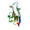
- PDB-1sjx: Three-Dimensional Structure of a Llama VHH Domain OE7 binding the... -
+ Open data

- Basic information
| Entry | Database: PDB / ID: 1sjx | ||||||
|---|---|---|---|---|---|---|---|
| Title | Three-Dimensional Structure of a Llama VHH Domain OE7 binding the cell wall protein Malf1 | ||||||
 Components Components | immunoglobulin VH domain | ||||||
 Keywords Keywords | IMMUNE SYSTEM / Camelids antibody / heavy chain / dandruff / phage display | ||||||
| Function / homology | Immunoglobulins / Immunoglobulin-like / Sandwich / Mainly Beta Function and homology information Function and homology information | ||||||
| Biological species |  | ||||||
| Method |  X-RAY DIFFRACTION / SYNCHROTRON / MOLECULAR REPLACEMENT / Resolution: 2.2 Å X-RAY DIFFRACTION / SYNCHROTRON / MOLECULAR REPLACEMENT / Resolution: 2.2 Å | ||||||
 Authors Authors | Dolk, E. / van der Vaart, M. / Hulsik, D.L. / Vriend, G. / de Haard, H. / Spinelli, S. / Cambillau, C. / Frenken, L. / Verrips, T. | ||||||
 Citation Citation | Journal: Appl.Environ.Microbiol. / Year: 2005 Title: Isolation of llama antibody fragments for prevention of dandruff by phage display in shampoo. Authors: Dolk, E. / van der Vaart, M. / Hulsik, D.L. / Vriend, G. / de Haard, H. / Spinelli, S. / Cambillau, C. / Frenken, L. / Verrips, T. | ||||||
| History |
| ||||||
| Remark 999 | SEQUENCE The sequence of the protein was not deposited into any sequence database. |
- Structure visualization
| Structure viewer | Molecule: MolmilJmol/JSmol |
|---|
- Downloads & links
-Download
| PDBx/mmCIF format | 1sjx.cif.gz | 38.8 KB | Display | PDBx/mmCIF format |
|---|---|---|---|---|
| PDB format | pdb1sjx.ent.gz | 26.2 KB | Display | PDB format |
| PDBx/mmJSON format | 1sjx.json.gz | Tree view | PDBx/mmJSON format | |
| Others |  Other downloads Other downloads |
-Validation report
| Arichive directory | https://data.pdbj.org/pub/pdb/validation_reports/sj/1sjxftp://data.pdbj.org/pub/pdb/validation_reports/sj/1sjx | HTTPS FTP |
|---|
-Related structure data
| Related structure data |  1hcvS S: Starting model for refinement |
|---|---|
| Similar structure data |


-Links
 PDBj
PDBj

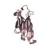
- Assembly
| Deposited unit | 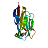
| ||||||||
|---|---|---|---|---|---|---|---|---|---|
| 1 |
| ||||||||
| Unit cell |
|
-Components
| #1: Antibody | Mass: 13214.672 Da / Num. of mol.: 1 Source method: isolated from a genetically manipulated source Source: (gene. exp.)  | ||||
|---|---|---|---|---|---|
| #2: Chemical |   Mass: 118.174 Da / Num. of mol.: 2 / Source method: obtained synthetically / Formula: C6H14O2 / Comment: precipitant*YM Mass: 118.174 Da / Num. of mol.: 2 / Source method: obtained synthetically / Formula: C6H14O2 / Comment: precipitant*YM#3: Water | ChemComp-HOH / |  Mass: 18.015 Da / Num. of mol.: 86 / Source method: isolated from a natural source / Formula: H2O Mass: 18.015 Da / Num. of mol.: 86 / Source method: isolated from a natural source / Formula: H2OHas protein modification | Y | |
-Experimental details
-Experiment
| Experiment | Method: X-RAY DIFFRACTION / Number of used crystals: 1 |
|---|
- Sample preparation
| Crystal | Density Matthews: 4.17 Å3/Da / Density % sol: 70.48 % |
|---|---|
| Crystal grow | Temperature: 293 K / Method: vapor diffusion, hanging drop / pH: 7.3 Details: 10% PEG 6K, 100mM Hepes, 5% MPD, pH 7.3, VAPOR DIFFUSION, HANGING DROP, temperature 293K |
-Data collection
| Diffraction | Mean temperature: 100 K |
|---|---|
| Diffraction source | Source: SYNCHROTRON / Site: ESRF  / Beamline: ID14-2 / Wavelength: 0.9326 Å / Beamline: ID14-2 / Wavelength: 0.9326 Å |
| Detector | Type: ADSC QUANTUM 4 / Detector: CCD / Date: Jun 3, 2000 |
| Radiation | Protocol: SINGLE WAVELENGTH / Monochromatic (M) / Laue (L): M / Scattering type: x-ray |
| Radiation wavelength | Wavelength: 0.9326 Å / Relative weight: 1 |
| Reflection | Resolution: 2.2→24 Å / Num. obs: 11594 / % possible obs: 99.8 % / Observed criterion σ(F): 3.5 / Observed criterion σ(I): 3.5 / Rmerge(I) obs: 0.042 / Rsym value: 0.042 / Net I/σ(I): 3.5 |
| Reflection shell | Resolution: 2.2→2.32 Å / Redundancy: 6 % / Rmerge(I) obs: 0.2 / Mean I/σ(I) obs: 3.5 / Num. unique all: 1637 / Rsym value: 0.2 / % possible all: 99.8 |
- Processing
| Software |
| ||||||||||||||||||||||||||||||||||||||||||||||||||||||||||||||||||||||||||||||||||||||||||||||||||||||||||||||||||||||||||||||||||||||||||||||||||||||||||||||||
|---|---|---|---|---|---|---|---|---|---|---|---|---|---|---|---|---|---|---|---|---|---|---|---|---|---|---|---|---|---|---|---|---|---|---|---|---|---|---|---|---|---|---|---|---|---|---|---|---|---|---|---|---|---|---|---|---|---|---|---|---|---|---|---|---|---|---|---|---|---|---|---|---|---|---|---|---|---|---|---|---|---|---|---|---|---|---|---|---|---|---|---|---|---|---|---|---|---|---|---|---|---|---|---|---|---|---|---|---|---|---|---|---|---|---|---|---|---|---|---|---|---|---|---|---|---|---|---|---|---|---|---|---|---|---|---|---|---|---|---|---|---|---|---|---|---|---|---|---|---|---|---|---|---|---|---|---|---|---|---|---|---|
| Refinement | Method to determine structure: MOLECULAR REPLACEMENT Starting model: PDB entry 1HCV Resolution: 2.2→24 Å / Cor.coef. Fo:Fc: 0.95 / Cor.coef. Fo:Fc free: 0.943 / SU B: 3.613 / SU ML: 0.092 / TLS residual ADP flag: LIKELY RESIDUAL / Cross valid method: THROUGHOUT / σ(F): 2 / ESU R: 0.165 / ESU R Free: 0.142 / Stereochemistry target values: MAXIMUM LIKELIHOOD
| ||||||||||||||||||||||||||||||||||||||||||||||||||||||||||||||||||||||||||||||||||||||||||||||||||||||||||||||||||||||||||||||||||||||||||||||||||||||||||||||||
| Solvent computation | Ion probe radii: 0.8 Å / Shrinkage radii: 0.8 Å / VDW probe radii: 1.4 Å / Solvent model: BABINET MODEL WITH MASK | ||||||||||||||||||||||||||||||||||||||||||||||||||||||||||||||||||||||||||||||||||||||||||||||||||||||||||||||||||||||||||||||||||||||||||||||||||||||||||||||||
| Displacement parameters | Biso mean: 22.713 Å2 | ||||||||||||||||||||||||||||||||||||||||||||||||||||||||||||||||||||||||||||||||||||||||||||||||||||||||||||||||||||||||||||||||||||||||||||||||||||||||||||||||
| Refinement step | Cycle: LAST / Resolution: 2.2→24 Å
| ||||||||||||||||||||||||||||||||||||||||||||||||||||||||||||||||||||||||||||||||||||||||||||||||||||||||||||||||||||||||||||||||||||||||||||||||||||||||||||||||
| Refine LS restraints |
| ||||||||||||||||||||||||||||||||||||||||||||||||||||||||||||||||||||||||||||||||||||||||||||||||||||||||||||||||||||||||||||||||||||||||||||||||||||||||||||||||
| LS refinement shell | Resolution: 2.199→2.256 Å / Total num. of bins used: 20 /
| ||||||||||||||||||||||||||||||||||||||||||||||||||||||||||||||||||||||||||||||||||||||||||||||||||||||||||||||||||||||||||||||||||||||||||||||||||||||||||||||||
| Refinement TLS params. | Method: refined / Origin x: -2.283 Å / Origin y: 49.94 Å / Origin z: 5.464 Å
|
