 Movie
Movie Controller

[English] 日本語
 Yorodumi
- PDB-1sh8: 1.5 A Crystal Structure of a Protein of Unknown Function PA5026 f... -
+ Open data

- Basic information
| Entry | Database: PDB / ID: 1sh8 | ||||||
|---|---|---|---|---|---|---|---|
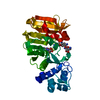
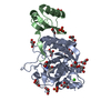
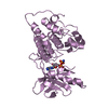
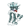
| Title | 1.5 A Crystal Structure of a Protein of Unknown Function PA5026 from Pseudomonas aeruginosa, Probable Thioesterase | ||||||
 Components Components | hypothetical protein PA5026 | ||||||
 Keywords Keywords | STRUCTURAL GENOMICS / UNKNOWN FUNCTION / Pseudomonas aeruginosa / PSI / Protein Structure Initiative / Midwest Center for Structural Genomics / MCSG | ||||||
| Function / homology | Protein of unknown function DUF4442 / Domain of unknown function (DUF4442) / Hotdog Thioesterase / Thiol Ester Dehydrase; Chain A / HotDog domain superfamily / Roll / Alpha Beta / DUF4442 domain-containing protein Function and homology information Function and homology information | ||||||
| Biological species |   Pseudomonas aeruginosa (bacteria) Pseudomonas aeruginosa (bacteria) | ||||||
| Method |  X-RAY DIFFRACTION / SYNCHROTRON / MAD / Resolution: 1.5 Å X-RAY DIFFRACTION / SYNCHROTRON / MAD / Resolution: 1.5 Å | ||||||
 Authors Authors | Zhang, R. / Evdokimova, E. / Savchenko, A. / Edwards, A. / Joachimiak, A. / Midwest Center for Structural Genomics (MCSG) | ||||||
 Citation Citation | Journal: To be Published Title: 1.5A crystal structure of a hypothetical protein PA5026 from Pseudomonas aeruginosa Authors: Zhang, R. / Evdokimova, E. / Savchenko, A. / Edwards, A. / Joachimiak, A. | ||||||
| History |
|
- Structure visualization
| Structure viewer | Molecule: MolmilJmol/JSmol |
|---|
- Downloads & links
-Download
| PDBx/mmCIF format | 1sh8.cif.gz | 76.5 KB | Display | PDBx/mmCIF format |
|---|---|---|---|---|
| PDB format | pdb1sh8.ent.gz | 57.6 KB | Display | PDB format |
| PDBx/mmJSON format | 1sh8.json.gz | Tree view | PDBx/mmJSON format | |
| Others |  Other downloads Other downloads |
-Validation report
| Arichive directory | https://data.pdbj.org/pub/pdb/validation_reports/sh/1sh8ftp://data.pdbj.org/pub/pdb/validation_reports/sh/1sh8 | HTTPS FTP |
|---|
-Related structure data
| Similar structure data | |
|---|---|
| Other databases |


-Links
 PDBj
PDBj
- Assembly
| Deposited unit | 
| ||||||||
|---|---|---|---|---|---|---|---|---|---|
| 1 |
| ||||||||
| Unit cell |
| ||||||||
| Details | This protein exists as dimer. Mol.A and Mol.B represents the dimer in asymetric unit. |
-Components
| #1: Protein | Mass: 17148.473 Da / Num. of mol.: 2 Source method: isolated from a genetically manipulated source Source: (gene. exp.) Pseudomonas aeruginosa (bacteria) / Strain: PA5026 / Plasmid: pET15b / Species (production host): Escherichia coli / Production host: Keywords: a few residues of histag plus protein plus 2 extra residues at C-terminal in mol.AReferences: UniProt: Q9HUE3 #2: Water | ChemComp-HOH / |  Mass: 18.015 Da / Num. of mol.: 332 / Source method: isolated from a natural source / Formula: H2O Mass: 18.015 Da / Num. of mol.: 332 / Source method: isolated from a natural source / Formula: H2O |
|---|
-Experimental details
-Experiment
| Experiment | Method: X-RAY DIFFRACTION / Number of used crystals: 1 |
|---|
- Sample preparation
| Crystal | Density Matthews: 2.8 Å3/Da / Density % sol: 55.73 % |
|---|---|
| Crystal grow | Temperature: 298 K / Method: vapor diffusion, hanging drop / pH: 4.6 Details: NaForm 2M, 0.1M NaAcet. 2%MPD, pH 4.6, VAPOR DIFFUSION, HANGING DROP, temperature 298K |
-Data collection
| Diffraction | Mean temperature: 100 K | ||||||||||||
|---|---|---|---|---|---|---|---|---|---|---|---|---|---|
| Diffraction source | Source: SYNCHROTRON / Site: APS  / Beamline: 19-ID / Wavelength: 0.9795,0.9797,0.94656 / Beamline: 19-ID / Wavelength: 0.9795,0.9797,0.94656 | ||||||||||||
| Detector | Type: SBC-2 / Detector: CCD / Date: Feb 8, 2004 / Details: morrors | ||||||||||||
| Radiation | Monochromator: Si 111 channel / Protocol: MAD / Monochromatic (M) / Laue (L): M / Scattering type: x-ray | ||||||||||||
| Radiation wavelength |
| ||||||||||||
| Reflection | Resolution: 1.5→50 Å / Num. all: 115037 / Num. obs: 109285 / % possible obs: 95 % / Observed criterion σ(F): 4 / Observed criterion σ(I): 4 / Redundancy: 4.97 % / Biso Wilson estimate: 13.3 Å2 / Rmerge(I) obs: 0.065 / Net I/σ(I): 21.25 | ||||||||||||
| Reflection shell | Resolution: 1.5→1.55 Å / Redundancy: 2.8 % / Rmerge(I) obs: 0.387 / Mean I/σ(I) obs: 1.98 / Num. unique all: 11501 / % possible all: 64.4 |
- Processing
| Software |
| |||||||||||||||||||||||||
|---|---|---|---|---|---|---|---|---|---|---|---|---|---|---|---|---|---|---|---|---|---|---|---|---|---|---|
| Refinement | Method to determine structure: MAD / Resolution: 1.5→23.24 Å / Rfactor Rfree error: 0.003 / Data cutoff high absF: 259680.66 / Data cutoff low absF: 0 / Isotropic thermal model: RESTRAINED / Cross valid method: THROUGHOUT / σ(F): 0 / Stereochemistry target values: Engh & Huber
| |||||||||||||||||||||||||
| Solvent computation | Solvent model: FLAT MODEL / Bsol: 54.3313 Å2 / ksol: 0.369007 e/Å3 | |||||||||||||||||||||||||
| Displacement parameters | Biso mean: 18.2 Å2
| |||||||||||||||||||||||||
| Refine analyze |
| |||||||||||||||||||||||||
| Refinement step | Cycle: LAST / Resolution: 1.5→23.24 Å
| |||||||||||||||||||||||||
| Refine LS restraints |
| |||||||||||||||||||||||||
| LS refinement shell | Resolution: 1.5→1.59 Å / Rfactor Rfree error: 0.012 / Total num. of bins used: 6
| |||||||||||||||||||||||||
| Xplor file |
|
