 Movie
Movie Controller

[English] 日本語
 Yorodumi
- PDB-1sc1: Crystal structure of an active-site ligand-free form of the human... -
+ Open data

- Basic information
| Entry | Database: PDB / ID: 1sc1 | ||||||
|---|---|---|---|---|---|---|---|
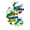
| Title | Crystal structure of an active-site ligand-free form of the human caspase-1 C285A mutant | ||||||
 Components Components | (Interleukin-1 beta convertase) x 2 | ||||||
 Keywords Keywords | HYDROLASE / ligand-free caspase-1 | ||||||
| Function / homology |  Function and homology information Function and homology informationcaspase-1 / protease inhibitor complex / AIM2 inflammasome complex assembly / IPAF inflammasome complex / The AIM2 inflammasome / AIM2 inflammasome complex / The IPAF inflammasome / icosanoid biosynthetic process / NLRP1 inflammasome complex / canonical inflammasome complex ...caspase-1 / protease inhibitor complex / AIM2 inflammasome complex assembly / IPAF inflammasome complex / The AIM2 inflammasome / AIM2 inflammasome complex / The IPAF inflammasome / icosanoid biosynthetic process / NLRP1 inflammasome complex / canonical inflammasome complex / positive regulation of interleukin-18 production / CARD domain binding / cytokine precursor processing / NLRP3 inflammasome complex / Interleukin-1 processing / Interleukin-37 signaling / positive regulation of tumor necrosis factor-mediated signaling pathway / osmosensory signaling pathway / signaling receptor ligand precursor processing / pattern recognition receptor signaling pathway / cysteine-type endopeptidase activator activity involved in apoptotic process / TP53 Regulates Transcription of Caspase Activators and Caspases / cytokine binding / protein autoprocessing / The NLRP3 inflammasome / pyroptotic inflammatory response / Pyroptosis / Purinergic signaling in leishmaniasis infection / positive regulation of interleukin-1 beta production / cellular response to mechanical stimulus / protein maturation / NOD1/2 Signaling Pathway / cellular response to type II interferon / kinase binding / SARS-CoV-1 activates/modulates innate immune responses / positive regulation of inflammatory response / cellular response to lipopolysaccharide / regulation of inflammatory response / endopeptidase activity / regulation of apoptotic process / defense response to virus / microtubule / positive regulation of canonical NF-kappaB signal transduction / defense response to bacterium / cysteine-type endopeptidase activity / apoptotic process / nucleolus / signal transduction / protein-containing complex / proteolysis / identical protein binding / plasma membrane / cytosol / cytoplasm Similarity search - Function | ||||||
| Biological species |  Homo sapiens (human) Homo sapiens (human) | ||||||
| Method |  X-RAY DIFFRACTION / MOLECULAR REPLACEMENT / Resolution: 2.6 Å X-RAY DIFFRACTION / MOLECULAR REPLACEMENT / Resolution: 2.6 Å | ||||||
 Authors Authors | Romanowski, M.J. / Scheer, J.M. / O'Brien, T. / McDowell, R.S. | ||||||
 Citation Citation | Journal: Structure / Year: 2004 Title: Crystal structures of a ligand-free and malonate-bound human caspase-1: implications for the mechanism of substrate binding. Authors: Romanowski, M.J. / Scheer, J.M. / O'Brien, T. / McDowell, R.S. | ||||||
| History |
|
- Structure visualization
| Structure viewer | Molecule: MolmilJmol/JSmol |
|---|
- Downloads & links
-Download
| PDBx/mmCIF format | 1sc1.cif.gz | 65.9 KB | Display | PDBx/mmCIF format |
|---|---|---|---|---|
| PDB format | pdb1sc1.ent.gz | 48.1 KB | Display | PDB format |
| PDBx/mmJSON format | 1sc1.json.gz | Tree view | PDBx/mmJSON format | |
| Others |  Other downloads Other downloads |
-Validation report
| Arichive directory | https://data.pdbj.org/pub/pdb/validation_reports/sc/1sc1ftp://data.pdbj.org/pub/pdb/validation_reports/sc/1sc1 | HTTPS FTP |
|---|
-Related structure data
| Related structure data |  1sc3C  1sc4C  1rwnS C: citing same article ( S: Starting model for refinement |
|---|---|
| Similar structure data |


-Links
 PDBj
PDBj


- Assembly
| Deposited unit | 
| ||||||||
|---|---|---|---|---|---|---|---|---|---|
| 1 | 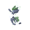
| ||||||||
| Unit cell |
|
-Components
| #1: Protein | Mass: 19837.773 Da / Num. of mol.: 1 / Fragment: INTERLEUKIN-1 BETA CONVERTASE P20 / Mutation: C285A Source method: isolated from a genetically manipulated source Source: (gene. exp.) Homo sapiens (human) / Gene: CASP1, IL1BC, IL1BCE / Plasmid: pRSET / Production host:  |
|---|---|
| #2: Protein | Mass: 10258.755 Da / Num. of mol.: 1 / Fragment: INTERLEUKIN-1 BETA CONVERTASE P10 Source method: isolated from a genetically manipulated source Source: (gene. exp.) Homo sapiens (human) / Gene: CASP1, IL1BC, IL1BCE / Plasmid: pRSET / Production host: |
| #3: Chemical | ChemComp-CL /   Mass: 35.453 Da / Num. of mol.: 1 / Source method: obtained synthetically / Formula: Cl Mass: 35.453 Da / Num. of mol.: 1 / Source method: obtained synthetically / Formula: Cl |
| #4: Water | ChemComp-HOH /  Mass: 18.015 Da / Num. of mol.: 60 / Source method: isolated from a natural source / Formula: H2O Mass: 18.015 Da / Num. of mol.: 60 / Source method: isolated from a natural source / Formula: H2O |
-Experimental details
-Experiment
| Experiment | Method: X-RAY DIFFRACTION / Number of used crystals: 1 |
|---|
- Sample preparation
| Crystal | Density Matthews: 2.93 Å3/Da / Density % sol: 57.96 % |
|---|---|
| Crystal grow | Temperature: 278 K / Method: vapor diffusion, hanging drop / pH: 7.4 Details: 0.1 M HEPES, 2 M (NH4)2SO4, 25 mM DTT. 0.01% Triton-X was added to the drop to prevent the crystals from attaching themselves to the cover slips., pH 7.4, VAPOR DIFFUSION, HANGING DROP, temperature 278K |
-Data collection
| Diffraction | Mean temperature: 180 K |
|---|---|
| Diffraction source | Source: ROTATING ANODE / Type: RIGAKU RUH3R / Wavelength: 1.5418 |
| Detector | Type: RIGAKU RAXIS IV / Detector: IMAGE PLATE / Date: May 27, 2003 |
| Radiation | Protocol: SINGLE WAVELENGTH / Monochromatic (M) / Laue (L): M / Scattering type: x-ray |
| Radiation wavelength | Wavelength: 1.5418 Å / Relative weight: 1 |
| Reflection | Resolution: 2.6→20 Å / Num. obs: 11308 / % possible obs: 99.6 % / Observed criterion σ(F): 0 / Observed criterion σ(I): 0 / Rmerge(I) obs: 0.107 |
| Reflection shell | Resolution: 2.6→2.69 Å / Rmerge(I) obs: 0.328 / % possible all: 100 |
- Processing
| Software |
| ||||||||||||||||||||||||||||||||||||||||||||||||||||||||||||||||||||||||||||||||||||||||||||||||||||||||||||||||||||||||||||||||||||||||||||||||||||||||||||||||
|---|---|---|---|---|---|---|---|---|---|---|---|---|---|---|---|---|---|---|---|---|---|---|---|---|---|---|---|---|---|---|---|---|---|---|---|---|---|---|---|---|---|---|---|---|---|---|---|---|---|---|---|---|---|---|---|---|---|---|---|---|---|---|---|---|---|---|---|---|---|---|---|---|---|---|---|---|---|---|---|---|---|---|---|---|---|---|---|---|---|---|---|---|---|---|---|---|---|---|---|---|---|---|---|---|---|---|---|---|---|---|---|---|---|---|---|---|---|---|---|---|---|---|---|---|---|---|---|---|---|---|---|---|---|---|---|---|---|---|---|---|---|---|---|---|---|---|---|---|---|---|---|---|---|---|---|---|---|---|---|---|---|
| Refinement | Method to determine structure: MOLECULAR REPLACEMENT Starting model: PDB ENTRY 1RWN Resolution: 2.6→20 Å / Cor.coef. Fo:Fc: 0.934 / Cor.coef. Fo:Fc free: 0.911 / SU B: 14.997 / SU ML: 0.29 / Cross valid method: THROUGHOUT / σ(F): 0 / σ(I): 0 / ESU R: 0.67 / ESU R Free: 0.333 / Stereochemistry target values: MAXIMUM LIKELIHOOD
| ||||||||||||||||||||||||||||||||||||||||||||||||||||||||||||||||||||||||||||||||||||||||||||||||||||||||||||||||||||||||||||||||||||||||||||||||||||||||||||||||
| Solvent computation | Ion probe radii: 0.8 Å / Shrinkage radii: 0.8 Å / VDW probe radii: 1.4 Å / Solvent model: MASK | ||||||||||||||||||||||||||||||||||||||||||||||||||||||||||||||||||||||||||||||||||||||||||||||||||||||||||||||||||||||||||||||||||||||||||||||||||||||||||||||||
| Displacement parameters | Biso mean: 51.343 Å2
| ||||||||||||||||||||||||||||||||||||||||||||||||||||||||||||||||||||||||||||||||||||||||||||||||||||||||||||||||||||||||||||||||||||||||||||||||||||||||||||||||
| Refinement step | Cycle: LAST / Resolution: 2.6→20 Å
| ||||||||||||||||||||||||||||||||||||||||||||||||||||||||||||||||||||||||||||||||||||||||||||||||||||||||||||||||||||||||||||||||||||||||||||||||||||||||||||||||
| Refine LS restraints |
| ||||||||||||||||||||||||||||||||||||||||||||||||||||||||||||||||||||||||||||||||||||||||||||||||||||||||||||||||||||||||||||||||||||||||||||||||||||||||||||||||
| LS refinement shell | Resolution: 2.6→2.69 Å / Total num. of bins used: 15 /
|
