 Movie
Movie Controller


+ Open data

- Basic information
| Entry | Database: PDB / ID: 1sav | ||||||
|---|---|---|---|---|---|---|---|
| Title | HUMAN ANNEXIN V WITH PROLINE SUBSTITUTION BY THIOPROLINE | ||||||
 Components Components | ANNEXIN V | ||||||
 Keywords Keywords | CALCIUM/PHOSPHOLIPID BINDING / THIOPROLINE / CALCIUM-PHOSPHOLIPID BINDING complex | ||||||
| Function / homology |  Function and homology information Function and homology informationphospholipase inhibitor activity / endothelial microparticle / negative regulation of coagulation / calcium-dependent phospholipid binding / vesicle membrane / phosphatidylserine binding / sarcolemma / phospholipid binding / blood coagulation / Platelet degranulation ...phospholipase inhibitor activity / endothelial microparticle / negative regulation of coagulation / calcium-dependent phospholipid binding / vesicle membrane / phosphatidylserine binding / sarcolemma / phospholipid binding / blood coagulation / Platelet degranulation / extracellular matrix / external side of plasma membrane / focal adhesion / calcium ion binding / negative regulation of apoptotic process / signal transduction / extracellular exosome / extracellular region / membrane / cytoplasm / cytosol Similarity search - Function | ||||||
| Biological species |  Homo sapiens (human) Homo sapiens (human) | ||||||
| Method |  X-RAY DIFFRACTION / MOLECULAR REPLACEMENT / Resolution: 2.5 Å X-RAY DIFFRACTION / MOLECULAR REPLACEMENT / Resolution: 2.5 Å | ||||||
 Authors Authors | Medrano, F.J. / Minks, C. / Budisa, N. / Huber, R. | ||||||
 Citation Citation | Journal: J.Mol.Biol. / Year: 1992 Title: Crystal and molecular structure of human annexin V after refinement. Implications for structure, membrane binding and ion channel formation of the annexin family of proteins. Authors: Huber, R. / Berendes, R. / Burger, A. / Schneider, M. / Karshikov, A. / Luecke, H. / Romisch, J. / Paques, E. #1: Journal: Embo J. / Year: 1990Title: The Crystal and Molecular Structure of Human Annexin V, an Anticoagulant Protein that Binds to Calcium and Membranes Authors: Huber, R. / Romisch, J. / Paques, E.P. #2: Journal: FEBS Lett. / Year: 1990Title: The Calcium Binding Sites in Human Annexin V by Crystal Structure Analysis at 2.0 A Resolution. Implications for Membrane Binding and Calcium Channel Activity Authors: Huber, R. / Schneider, M. / Mayr, I. / Romisch, J. / Paques, E.P. | ||||||
| History |
|
- Structure visualization
| Structure viewer | Molecule: MolmilJmol/JSmol |
|---|
- Downloads & links
-Download
| PDBx/mmCIF format | 1sav.cif.gz | 78.9 KB | Display | PDBx/mmCIF format |
|---|---|---|---|---|
| PDB format | pdb1sav.ent.gz | 58.9 KB | Display | PDB format |
| PDBx/mmJSON format | 1sav.json.gz | Tree view | PDBx/mmJSON format | |
| Others |  Other downloads Other downloads |
-Validation report
| Arichive directory | https://data.pdbj.org/pub/pdb/validation_reports/sa/1savftp://data.pdbj.org/pub/pdb/validation_reports/sa/1sav | HTTPS FTP |
|---|
-Related structure data
| Related structure data | 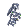 1avhC 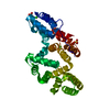 1avrSC S: Starting model for refinement C: citing same article ( |
|---|---|
| Similar structure data |


-Links
 PDBj
PDBj- Assembly
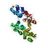
| Deposited unit | 
| ||||||||
|---|---|---|---|---|---|---|---|---|---|
| 1 |
| ||||||||
| Unit cell |
|
-Components
| #1: Protein | Mass: 36070.902 Da / Num. of mol.: 1 Mutation: P13, P87, P119, P163, AND P248 SUBSTITUTED WITH THIOPROLINE (PRS) Source method: isolated from a genetically manipulated source Source: (gene. exp.) Homo sapiens (human) / Production host:  | ||||
|---|---|---|---|---|---|
| #2: Chemical | ChemComp-CA /   Mass: 40.078 Da / Num. of mol.: 5 / Source method: obtained synthetically / Formula: Ca Mass: 40.078 Da / Num. of mol.: 5 / Source method: obtained synthetically / Formula: Ca#3: Water | ChemComp-HOH / |  Mass: 18.015 Da / Num. of mol.: 152 / Source method: isolated from a natural source / Formula: H2O Mass: 18.015 Da / Num. of mol.: 152 / Source method: isolated from a natural source / Formula: H2OHas protein modification | Y | |
-Experimental details
-Experiment
| Experiment | Method: X-RAY DIFFRACTION / Number of used crystals: 1 |
|---|
- Sample preparation
| Crystal | Density Matthews: 2.57 Å3/Da / Density % sol: 52.1 % | |||||||||||||||||||||||||||||||||||||||||||||||||
|---|---|---|---|---|---|---|---|---|---|---|---|---|---|---|---|---|---|---|---|---|---|---|---|---|---|---|---|---|---|---|---|---|---|---|---|---|---|---|---|---|---|---|---|---|---|---|---|---|---|---|
| Crystal grow | pH: 8.5 Details: PROTEIN WAS CRYSTALLIZED FROM 2.1 M AMMONIUM SULFATE, 0.1 M TRIS-HCL, PH 8.5 | |||||||||||||||||||||||||||||||||||||||||||||||||
| Crystal grow | *PLUS pH: 7 / Method: vapor diffusion | |||||||||||||||||||||||||||||||||||||||||||||||||
| Components of the solutions | *PLUS
|
-Data collection
| Diffraction | Mean temperature: 280 K |
|---|---|
| Diffraction source | Source: ROTATING ANODE / Type: RIGAKU RUH2R / Wavelength: 1.5418 |
| Detector | Type: MARRESEARCH / Detector: IMAGE PLATE / Date: Feb 1, 1997 |
| Radiation | Monochromator: GRAPHITE(002) / Monochromatic (M) / Laue (L): M / Scattering type: x-ray |
| Radiation wavelength | Wavelength: 1.5418 Å / Relative weight: 1 |
| Reflection | Resolution: 2.5→25 Å / Num. obs: 12532 / % possible obs: 99.3 % / Observed criterion σ(I): 2 / Redundancy: 4.5 % / Rmerge(I) obs: 0.049 / Net I/σ(I): 9.5 |
| Reflection shell | Resolution: 2.5→2.55 Å / % possible all: 95.8 |
- Processing
| Software |
| ||||||||||||||||||||||||||||||||||||||||||||||||||||||||||||
|---|---|---|---|---|---|---|---|---|---|---|---|---|---|---|---|---|---|---|---|---|---|---|---|---|---|---|---|---|---|---|---|---|---|---|---|---|---|---|---|---|---|---|---|---|---|---|---|---|---|---|---|---|---|---|---|---|---|---|---|---|---|
| Refinement | Method to determine structure: MOLECULAR REPLACEMENT Starting model: PDB ENTRY 1AVR Resolution: 2.5→8 Å / Data cutoff high absF: 1000000 / Data cutoff low absF: 0.001 / Cross valid method: THROUGHOUT / σ(F): 2
| ||||||||||||||||||||||||||||||||||||||||||||||||||||||||||||
| Refinement step | Cycle: LAST / Resolution: 2.5→8 Å
| ||||||||||||||||||||||||||||||||||||||||||||||||||||||||||||
| Refine LS restraints |
|
