 Movie
Movie Controller

[English] 日本語
 Yorodumi
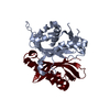
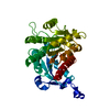
- PDB-1s7i: 1.8 A Crystal Structure of a Protein of Unknown Function PA1349 f... -
+ Open data

- Basic information
| Entry | Database: PDB / ID: 1s7i | ||||||
|---|---|---|---|---|---|---|---|
| Title | 1.8 A Crystal Structure of a Protein of Unknown Function PA1349 from Pseudomonas aeruginosa | ||||||
 Components Components | hypothetical protein PA1349 | ||||||
 Keywords Keywords | STRUCTURAL GENOMICS / UNKNOWN FUNCTION / protein structure initiative / Pseudomonas aeruginosa / MCSG / PSI / Midwest Center for Structural Genomics | ||||||
| Function / homology | YCII-related / YCII-related domain / Dimeric alpha+beta barrel / Dimeric alpha-beta barrel / Alpha-Beta Plaits / 2-Layer Sandwich / Alpha Beta / YCII-related domain-containing protein Function and homology information Function and homology information | ||||||
| Biological species |   Pseudomonas aeruginosa (bacteria) Pseudomonas aeruginosa (bacteria) | ||||||
| Method |  X-RAY DIFFRACTION / SYNCHROTRON / MAD / Resolution: 1.8 Å X-RAY DIFFRACTION / SYNCHROTRON / MAD / Resolution: 1.8 Å | ||||||
 Authors Authors | Zhang, R. / Skarina, T. / Savchenko, A. / Edwards, A. / Joachimiak, A. / Midwest Center for Structural Genomics (MCSG) | ||||||
 Citation Citation | Journal: To be Published Title: 1.8A crystal structure of a hypothetical protein PA1349 from Pseudomonas aeruginosa Authors: Zhang, R. / Skarina, T. / Savchenko, A. / Edwards, A. / Joachimiak, A. | ||||||
| History |
|
- Structure visualization
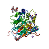
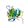
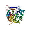
| Structure viewer | Molecule: MolmilJmol/JSmol |
|---|
- Downloads & links
-Download
| PDBx/mmCIF format | 1s7i.cif.gz | 36.9 KB | Display | PDBx/mmCIF format |
|---|---|---|---|---|
| PDB format | pdb1s7i.ent.gz | 25.7 KB | Display | PDB format |
| PDBx/mmJSON format | 1s7i.json.gz | Tree view | PDBx/mmJSON format | |
| Others |  Other downloads Other downloads |
-Validation report
| Arichive directory | https://data.pdbj.org/pub/pdb/validation_reports/s7/1s7iftp://data.pdbj.org/pub/pdb/validation_reports/s7/1s7i | HTTPS FTP |
|---|
-Related structure data
| Similar structure data | |
|---|---|
| Other databases |


-Links
 PDBj
PDBj- Assembly
| Deposited unit | 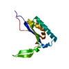
| ||||||||
|---|---|---|---|---|---|---|---|---|---|
| 1 | 
| ||||||||
| Unit cell |
| ||||||||
| Details | This protein existed as dimer. The second part of the biological assembly is generated by the crystallographic two fold axis |
-Components
| #1: Protein | Mass: 13633.543 Da / Num. of mol.: 1 Source method: isolated from a genetically manipulated source Source: (gene. exp.) Pseudomonas aeruginosa (bacteria) / Strain: PA1349 / Plasmid: pET15b / Species (production host): Escherichia coli / Production host: |
|---|---|
| #2: Water | ChemComp-HOH /  Mass: 18.015 Da / Num. of mol.: 57 / Source method: isolated from a natural source / Formula: H2O Mass: 18.015 Da / Num. of mol.: 57 / Source method: isolated from a natural source / Formula: H2O |
-Experimental details
-Experiment
| Experiment | Method: X-RAY DIFFRACTION / Number of used crystals: 1 |
|---|
- Sample preparation
| Crystal | Density Matthews: 3.44 Å3/Da / Density % sol: 63.98 % |
|---|---|
| Crystal grow | Temperature: 298 K / Method: vapor diffusion, hanging drop / pH: 8.5 Details: 1.5M Amm. Sulph., 0.1M Tris, pH 8.5, VAPOR DIFFUSION, HANGING DROP, temperature 298K |
-Data collection
| Diffraction | Mean temperature: 100 K | ||||||||||||
|---|---|---|---|---|---|---|---|---|---|---|---|---|---|
| Diffraction source | Source: SYNCHROTRON / Site: APS  / Beamline: 19-ID / Wavelength: 0.9795, 0.9797, 0.94656 / Beamline: 19-ID / Wavelength: 0.9795, 0.9797, 0.94656 | ||||||||||||
| Detector | Type: SBC-2 / Detector: CCD / Date: Dec 11, 2003 / Details: mirrors | ||||||||||||
| Radiation | Monochromator: Si 111 channel / Protocol: MAD / Monochromatic (M) / Laue (L): M / Scattering type: x-ray | ||||||||||||
| Radiation wavelength |
| ||||||||||||
| Reflection | Resolution: 1.8→50 Å / Num. all: 18121 / Num. obs: 17179 / % possible obs: 94.8 % / Observed criterion σ(F): 4 / Observed criterion σ(I): 4 / Redundancy: 9.12 % / Biso Wilson estimate: 22.6 Å2 / Rmerge(I) obs: 0.048 / Net I/σ(I): 41.33 | ||||||||||||
| Reflection shell | Resolution: 1.8→1.86 Å / Redundancy: 5.6 % / Rmerge(I) obs: 0.68 / Mean I/σ(I) obs: 1.71 / Num. unique all: 1759 / % possible all: 76.8 |
- Processing
| Software |
| ||||||||||||||||||||
|---|---|---|---|---|---|---|---|---|---|---|---|---|---|---|---|---|---|---|---|---|---|
| Refinement | Method to determine structure: MAD / Resolution: 1.8→28.69 Å / Rfactor Rfree error: 0.007 / Data cutoff high absF: 827357.77 / Data cutoff low absF: 0 / Isotropic thermal model: RESTRAINED / Cross valid method: THROUGHOUT / σ(F): 0 / Stereochemistry target values: Engh & Huber
| ||||||||||||||||||||
| Solvent computation | Solvent model: FLAT MODEL / Bsol: 56.9607 Å2 / ksol: 0.376487 e/Å3 | ||||||||||||||||||||
| Displacement parameters | Biso mean: 33.4 Å2
| ||||||||||||||||||||
| Refine analyze |
| ||||||||||||||||||||
| Refinement step | Cycle: LAST / Resolution: 1.8→28.69 Å
| ||||||||||||||||||||
| Refine LS restraints |
| ||||||||||||||||||||
| LS refinement shell | Resolution: 1.8→1.91 Å / Rfactor Rfree error: 0.021 / Total num. of bins used: 6
| ||||||||||||||||||||
| Xplor file |
|
