 Movie
Movie Controller


+ Open data

- Basic information
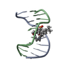
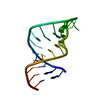
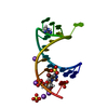
| Entry | Database: PDB / ID: 1s34 | ||||||||||||||||||
|---|---|---|---|---|---|---|---|---|---|---|---|---|---|---|---|---|---|---|---|
| Title | Solution structure of residues 907-929 from Rous Sarcoma Virus | ||||||||||||||||||
 Components Components | 5'-R(* Keywords KeywordsRNA / stem-loop / tetraloop / bulge | Function / homology | RNA / RNA (> 10) |  Function and homology information Function and homology informationMethod | SOLUTION NMR / Simulated annealing. Refinement using torsion angle dynamics, the DELPHIC database |  Authors AuthorsCabello-Villegas, J. / Giles, K.E. / Soto, A.M. / Yu, P. / Beemon, K.L. / Wang, Y.X. |  CitationJournal: Rna / Year: 2004 CitationJournal: Rna / Year: 2004Title: Solution structure of the pseudo-5' splice site of a retroviral splicing suppressor. Authors: Cabello-Villegas, J. / Giles, K.E. / Soto, A.M. / Yu, P. / Mougin, A. / Beemon, K.L. / Wang, Y.X. History |
|
- Structure visualization
| Structure viewer | Molecule: MolmilJmol/JSmol |
|---|
- Downloads & links
-Download
| PDBx/mmCIF format | 1s34.cif.gz | 213.4 KB | Display | PDBx/mmCIF format |
|---|---|---|---|---|
| PDB format | pdb1s34.ent.gz | 179.9 KB | Display | PDB format |
| PDBx/mmJSON format | 1s34.json.gz | Tree view | PDBx/mmJSON format | |
| Others |  Other downloads Other downloads |
-Validation report
| Arichive directory | https://data.pdbj.org/pub/pdb/validation_reports/s3/1s34ftp://data.pdbj.org/pub/pdb/validation_reports/s3/1s34 | HTTPS FTP |
|---|
-Related structure data


-Links
 PDBj
PDBj


- Assembly
| Deposited unit | 
| |||||||||
|---|---|---|---|---|---|---|---|---|---|---|
| 1 |
| |||||||||
| NMR ensembles |
|
-Components
| #1: RNA chain | Mass: 7350.335 Da / Num. of mol.: 1 / Fragment: Pseudo 5'-splice site / Source method: obtained synthetically / Details: In vitro transcription |
|---|
-Experimental details
-Experiment
| Experiment | Method: SOLUTION NMR | ||||||||||||||||||||||||||||
|---|---|---|---|---|---|---|---|---|---|---|---|---|---|---|---|---|---|---|---|---|---|---|---|---|---|---|---|---|---|
| NMR experiment |
| ||||||||||||||||||||||||||||
| NMR details | Text: RDC restraints were obtained for the base one bond H-C and H-N using HSQC experiments. RDC restraints for the ribose were obtained from measurements on an Relay HCCH-COSY (Vallurupalli and ...Text: RDC restraints were obtained for the base one bond H-C and H-N using HSQC experiments. RDC restraints for the ribose were obtained from measurements on an Relay HCCH-COSY (Vallurupalli and Moore, J Biomol NMR. 2002. 24(1):63-6). Anisotropic conditions were ~20 mg/ml pf1 phage. |
- Sample preparation
| Details |
| |||||||||||||||
|---|---|---|---|---|---|---|---|---|---|---|---|---|---|---|---|---|
| Sample conditions |
|
-NMR measurement
| Radiation | Protocol: SINGLE WAVELENGTH / Monochromatic (M) / Laue (L): M | |||||||||||||||||||||||||
|---|---|---|---|---|---|---|---|---|---|---|---|---|---|---|---|---|---|---|---|---|---|---|---|---|---|---|
| Radiation wavelength | Relative weight: 1 | |||||||||||||||||||||||||
| NMR spectrometer |
|
- Processing
| NMR software |
| ||||||||||||||||||||||||||||
|---|---|---|---|---|---|---|---|---|---|---|---|---|---|---|---|---|---|---|---|---|---|---|---|---|---|---|---|---|---|
| Refinement | Method: Simulated annealing. Refinement using torsion angle dynamics, the DELPHIC database Software ordinal: 1 Details: DELPHIC torsion angle potential was used for the whole molecule. The base position potential was excluded for residues 913, and 917 to 920. Protocol from Clore and Kuszewski (J Am Chem Soc. 2003. 125(6):1518-25) | ||||||||||||||||||||||||||||
| NMR representative | Selection criteria: lowest energy | ||||||||||||||||||||||||||||
| NMR ensemble | Conformer selection criteria: structures with the lowest energy Conformers calculated total number: 100 / Conformers submitted total number: 15 |
 NMRPipe