 Movie
Movie Controller

[English] 日本語
 Yorodumi
- PDB-1rxo: ACTIVATED SPINACH RUBISCO IN COMPLEX WITH ITS SUBSTRATE RIBULOSE-... -
+ Open data

- Basic information
| Entry | Database: PDB / ID: 1rxo | ||||||
|---|---|---|---|---|---|---|---|
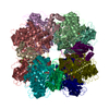
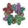
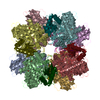
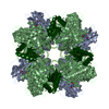
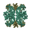
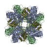
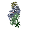
| Title | ACTIVATED SPINACH RUBISCO IN COMPLEX WITH ITS SUBSTRATE RIBULOSE-1,5-BISPHOSPHATE AND CALCIUM | ||||||
 Components Components | (RIBULOSE BISPHOSPHATE CARBOXYLASE/OXYGENASE) x 2 | ||||||
 Keywords Keywords | LYASE (CARBON-CARBON) | ||||||
| Function / homology |  Function and homology information Function and homology informationphotorespiration / ribulose-bisphosphate carboxylase / ribulose-bisphosphate carboxylase activity / reductive pentose-phosphate cycle / chloroplast / monooxygenase activity / magnesium ion binding Similarity search - Function | ||||||
| Biological species |  Spinacia oleracea (spinach) Spinacia oleracea (spinach) | ||||||
| Method |  X-RAY DIFFRACTION / SYNCHROTRON / ISOMORPHOUS WITH PDB ENTRY 1AUS / Resolution: 2.2 Å X-RAY DIFFRACTION / SYNCHROTRON / ISOMORPHOUS WITH PDB ENTRY 1AUS / Resolution: 2.2 Å | ||||||
 Authors Authors | Taylor, T.C. / Andersson, I. | ||||||
 Citation Citation | Journal: J.Mol.Biol. / Year: 1997 Title: The structure of the complex between rubisco and its natural substrate ribulose 1,5-bisphosphate. Authors: Taylor, T.C. / Andersson, I. #1: Journal: J.Mol.Biol. / Year: 1996Title: Large Structures at High Resolution: The 1.6 A Crystal Structure of Spinach Ribulose-1,5-Bisphosphate Carboxylase/Oxygenase Complexed with 2-Carboxyarabinitol Bisphosphate Authors: Andersson, I. | ||||||
| History |
|
- Structure visualization
| Structure viewer | Molecule: MolmilJmol/JSmol |
|---|
- Downloads & links
-Download
| PDBx/mmCIF format | 1rxo.cif.gz | 473.1 KB | Display | PDBx/mmCIF format |
|---|---|---|---|---|
| PDB format | pdb1rxo.ent.gz | 387.1 KB | Display | PDB format |
| PDBx/mmJSON format | 1rxo.json.gz | Tree view | PDBx/mmJSON format | |
| Others |  Other downloads Other downloads |
-Validation report
| Arichive directory | https://data.pdbj.org/pub/pdb/validation_reports/rx/1rxoftp://data.pdbj.org/pub/pdb/validation_reports/rx/1rxo | HTTPS FTP |
|---|
-Related structure data


-Links
 PDBj
PDBj
- Assembly
| Deposited unit | 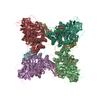
| ||||||||||||||||
|---|---|---|---|---|---|---|---|---|---|---|---|---|---|---|---|---|---|
| 1 | 
| ||||||||||||||||
| Unit cell |
| ||||||||||||||||
| Noncrystallographic symmetry (NCS) | NCS oper:
| ||||||||||||||||
| Details | THE DEPOSITORS PROVIDED CHAINS L AND S. THE OTHER CHAINS TO MAKE THE COMPLETE ASYMMETRIC UNIT WERE GENERATED BY THE PROTEIN DATA BANK USING THE MTRIX TRANSFORMATIONS BELOW. |
-Components
| #1: Protein | Mass: 52777.680 Da / Num. of mol.: 4 / Source method: isolated from a natural source / Source: (natural) Spinacia oleracea (spinach) / Organ: LEAFReferences: UniProt: P00875, ribulose-bisphosphate carboxylase #2: Protein | Mass: 14638.671 Da / Num. of mol.: 4 / Source method: isolated from a natural source / Source: (natural) Spinacia oleracea (spinach) / Organ: LEAFReferences: UniProt: P00870, ribulose-bisphosphate carboxylase #3: Sugar | ChemComp-RUB /   Type: saccharide / Mass: 310.090 Da / Num. of mol.: 4 Type: saccharide / Mass: 310.090 Da / Num. of mol.: 4Source method: isolated from a genetically manipulated source Formula: C5H12O11P2 #4: Chemical | ChemComp-CA /   Mass: 40.078 Da / Num. of mol.: 4 / Source method: obtained synthetically / Formula: Ca Mass: 40.078 Da / Num. of mol.: 4 / Source method: obtained synthetically / Formula: Ca#5: Water | ChemComp-HOH / |  Mass: 18.015 Da / Num. of mol.: 1024 / Source method: isolated from a natural source / Formula: H2O Mass: 18.015 Da / Num. of mol.: 1024 / Source method: isolated from a natural source / Formula: H2O |
|---|
-Experimental details
-Experiment
| Experiment | Method: X-RAY DIFFRACTION / Number of used crystals: 4 |
|---|
- Sample preparation
| Crystal | Density Matthews: 2.34 Å3/Da / Density % sol: 40 % | ||||||||||||||||||||||||||||||||||||||||||||||||||||||||||||||||||||||||||||||
|---|---|---|---|---|---|---|---|---|---|---|---|---|---|---|---|---|---|---|---|---|---|---|---|---|---|---|---|---|---|---|---|---|---|---|---|---|---|---|---|---|---|---|---|---|---|---|---|---|---|---|---|---|---|---|---|---|---|---|---|---|---|---|---|---|---|---|---|---|---|---|---|---|---|---|---|---|---|---|---|
| Crystal grow | pH: 7.8 Details: 14% PEG4000, 25 MM HEPES PH 7.8, 0.2 M NACL, 10 MM CACL2; SOAKED WITH 20 MM RUBP FOR 5 DAYS | ||||||||||||||||||||||||||||||||||||||||||||||||||||||||||||||||||||||||||||||
| Crystal | *PLUS | ||||||||||||||||||||||||||||||||||||||||||||||||||||||||||||||||||||||||||||||
| Crystal grow | *PLUS Temperature: 4 ℃ / Method: vapor diffusion, hanging drop / Details: Taylor, T.C., (1996) Nat. Struct. Biol., 3, 95. | ||||||||||||||||||||||||||||||||||||||||||||||||||||||||||||||||||||||||||||||
| Components of the solutions | *PLUS
|
-Data collection
| Diffraction | Mean temperature: 279 K |
|---|---|
| Diffraction source | Source: SYNCHROTRON / Site: SRS  / Beamline: PX9.6 / Wavelength: 0.87 / Beamline: PX9.6 / Wavelength: 0.87 |
| Detector | Type: MARRESEARCH / Detector: IMAGE PLATE / Date: Sep 1, 1994 |
| Radiation | Monochromatic (M) / Laue (L): M / Scattering type: x-ray |
| Radiation wavelength | Wavelength: 0.87 Å / Relative weight: 1 |
| Reflection | Resolution: 2.1→20 Å / Num. obs: 109900 / % possible obs: 75.5 % / Redundancy: 6 % / Rmerge(I) obs: 0.136 / Net I/σ(I): 6.9 |
| Reflection shell | Resolution: 2.1→2.17 Å / Redundancy: 2 % / Rmerge(I) obs: 0.247 / Mean I/σ(I) obs: 2.8 / % possible all: 53.8 |
| Reflection | *PLUS Num. measured all: 1275669 |
- Processing
| Software |
| ||||||||||||||||||||||||||||||||||||||||||||||||||||||||||||
|---|---|---|---|---|---|---|---|---|---|---|---|---|---|---|---|---|---|---|---|---|---|---|---|---|---|---|---|---|---|---|---|---|---|---|---|---|---|---|---|---|---|---|---|---|---|---|---|---|---|---|---|---|---|---|---|---|---|---|---|---|---|
| Refinement | Method to determine structure: ISOMORPHOUS WITH PDB ENTRY 1AUS Resolution: 2.2→7 Å / Isotropic thermal model: INDIVIDUAL / Cross valid method: THROUGHOUT / σ(F): 0
| ||||||||||||||||||||||||||||||||||||||||||||||||||||||||||||
| Refinement step | Cycle: LAST / Resolution: 2.2→7 Å
| ||||||||||||||||||||||||||||||||||||||||||||||||||||||||||||
| Refine LS restraints |
| ||||||||||||||||||||||||||||||||||||||||||||||||||||||||||||
| Refine LS restraints NCS | NCS model details: CONSTRAINTS | ||||||||||||||||||||||||||||||||||||||||||||||||||||||||||||
| LS refinement shell | Resolution: 2.2→2.3 Å / Total num. of bins used: 8
| ||||||||||||||||||||||||||||||||||||||||||||||||||||||||||||
| Xplor file |
| ||||||||||||||||||||||||||||||||||||||||||||||||||||||||||||
| Software | *PLUS Name: X-PLOR / Version: 3.1 / Classification: refinement | ||||||||||||||||||||||||||||||||||||||||||||||||||||||||||||
| Refine LS restraints | *PLUS
| ||||||||||||||||||||||||||||||||||||||||||||||||||||||||||||
| LS refinement shell | *PLUS Rfactor Rfree: 0.27 |
