 Movie
Movie Controller

[English] 日本語
 Yorodumi
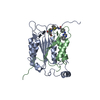
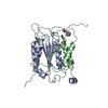
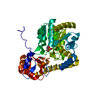
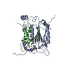
- PDB-1rwk: Crystal structure of human caspase-1 in complex with 3-(2-mercapt... -
+ Open data

- Basic information
| Entry | Database: PDB / ID: 1rwk | ||||||
|---|---|---|---|---|---|---|---|
| Title | Crystal structure of human caspase-1 in complex with 3-(2-mercapto-acetylamino)-4-oxo-pentanoic acid | ||||||
 Components Components | (Interleukin-1 beta convertase) x 2 | ||||||
 Keywords Keywords | HYDROLASE / protein-small molecule inhibitor complex | ||||||
| Function / homology |  Function and homology information Function and homology informationcaspase-1 / protease inhibitor complex / AIM2 inflammasome complex assembly / IPAF inflammasome complex / The AIM2 inflammasome / AIM2 inflammasome complex / The IPAF inflammasome / icosanoid biosynthetic process / NLRP1 inflammasome complex / canonical inflammasome complex ...caspase-1 / protease inhibitor complex / AIM2 inflammasome complex assembly / IPAF inflammasome complex / The AIM2 inflammasome / AIM2 inflammasome complex / The IPAF inflammasome / icosanoid biosynthetic process / NLRP1 inflammasome complex / canonical inflammasome complex / positive regulation of interleukin-18 production / CARD domain binding / cytokine precursor processing / NLRP3 inflammasome complex / Interleukin-1 processing / Interleukin-37 signaling / positive regulation of tumor necrosis factor-mediated signaling pathway / osmosensory signaling pathway / signaling receptor ligand precursor processing / pattern recognition receptor signaling pathway / cysteine-type endopeptidase activator activity involved in apoptotic process / TP53 Regulates Transcription of Caspase Activators and Caspases / cytokine binding / protein autoprocessing / The NLRP3 inflammasome / pyroptotic inflammatory response / Pyroptosis / Purinergic signaling in leishmaniasis infection / positive regulation of interleukin-1 beta production / cellular response to mechanical stimulus / protein maturation / NOD1/2 Signaling Pathway / cellular response to type II interferon / kinase binding / SARS-CoV-1 activates/modulates innate immune responses / positive regulation of inflammatory response / cellular response to lipopolysaccharide / regulation of inflammatory response / endopeptidase activity / regulation of apoptotic process / defense response to virus / microtubule / positive regulation of canonical NF-kappaB signal transduction / defense response to bacterium / cysteine-type endopeptidase activity / apoptotic process / nucleolus / signal transduction / protein-containing complex / proteolysis / identical protein binding / plasma membrane / cytosol / cytoplasm Similarity search - Function | ||||||
| Biological species |  Homo sapiens (human) Homo sapiens (human) | ||||||
| Method |  X-RAY DIFFRACTION / SYNCHROTRON / MOLECULAR REPLACEMENT / Resolution: 2.3 Å X-RAY DIFFRACTION / SYNCHROTRON / MOLECULAR REPLACEMENT / Resolution: 2.3 Å | ||||||
 Authors Authors | Romanowski, M.J. / Lam, J.W. / Fahr, B.T. / O'Brien, T. | ||||||
 Citation Citation | Journal: Acta Crystallogr.,Sect.F / Year: 2005 Title: Structural analysis of caspase-1 inhibitors derived from Tethering. Authors: O'Brien, T. / Fahr, B.T. / Sopko, M.M. / Lam, J.W. / Waal, N.D. / Raimundo, B.C. / Purkey, H.E. / Pham, P. / Romanowski, M.J. | ||||||
| History |
|
- Structure visualization
| Structure viewer | Molecule: MolmilJmol/JSmol |
|---|
- Downloads & links
-Download
| PDBx/mmCIF format | 1rwk.cif.gz | 69.2 KB | Display | PDBx/mmCIF format |
|---|---|---|---|---|
| PDB format | pdb1rwk.ent.gz | 51.1 KB | Display | PDB format |
| PDBx/mmJSON format | 1rwk.json.gz | Tree view | PDBx/mmJSON format | |
| Others |  Other downloads Other downloads |
-Validation report
| Arichive directory | https://data.pdbj.org/pub/pdb/validation_reports/rw/1rwkftp://data.pdbj.org/pub/pdb/validation_reports/rw/1rwk | HTTPS FTP |
|---|
-Related structure data
| Related structure data |  1rwmC  1rwnC  1rwoC  1rwpC  1iceS S: Starting model for refinement C: citing same article ( |
|---|---|
| Similar structure data |


-Links
 PDBj
PDBj


- Assembly
| Deposited unit | 
| ||||||||
|---|---|---|---|---|---|---|---|---|---|
| 1 | 
| ||||||||
| Unit cell |
| ||||||||
| Details | Heterotetramer (dimer of two heterodimers). Each heterodimer is represented by chains A (the p20 subunit) and B (the p10 subunit) of human caspase-1. |
-Components
| #1: Protein | Mass: 19869.838 Da / Num. of mol.: 1 / Fragment: INTERLEUKIN-1 BETA CONVERTASE P20 Source method: isolated from a genetically manipulated source Source: (gene. exp.) Homo sapiens (human) / Gene: CASP1, IL1BC, IL1BCE / Plasmid: pRSET / Production host:  |
|---|---|
| #2: Protein | Mass: 10258.755 Da / Num. of mol.: 1 / Fragment: INTERLEUKIN-1 BETA CONVERTASE P10 Source method: isolated from a genetically manipulated source Source: (gene. exp.) Homo sapiens (human) / Gene: CASP1, IL1BC, IL1BCE / Plasmid: pRSET / Production host: |
| #3: Chemical | ChemComp-158 /   Mass: 205.232 Da / Num. of mol.: 1 / Source method: obtained synthetically / Formula: C7H11NO4S Mass: 205.232 Da / Num. of mol.: 1 / Source method: obtained synthetically / Formula: C7H11NO4S |
| #4: Water | ChemComp-HOH /  Mass: 18.015 Da / Num. of mol.: 161 / Source method: isolated from a natural source / Formula: H2O Mass: 18.015 Da / Num. of mol.: 161 / Source method: isolated from a natural source / Formula: H2O |
| Has protein modification | Y |
-Experimental details
-Experiment
| Experiment | Method: X-RAY DIFFRACTION / Number of used crystals: 1 |
|---|
- Sample preparation
| Crystal | Density Matthews: 2.64 Å3/Da / Density % sol: 53.33 % |
|---|---|
| Crystal grow | Temperature: 274 K / Method: vapor diffusion, hanging drop / pH: 7 Details: 0.1M HEPES, 2M (NH4)2SO4, pH 7.0, VAPOR DIFFUSION, HANGING DROP, temperature 274K |
-Data collection
| Diffraction | Mean temperature: 180 K |
|---|---|
| Diffraction source | Source: SYNCHROTRON / Site: SSRL  / Beamline: BL7-1 / Wavelength: 0.98 Å / Beamline: BL7-1 / Wavelength: 0.98 Å |
| Detector | Detector: IMAGE PLATE |
| Radiation | Protocol: SINGLE WAVELENGTH / Monochromatic (M) / Laue (L): M / Scattering type: x-ray |
| Radiation wavelength | Wavelength: 0.98 Å / Relative weight: 1 |
| Reflection | Resolution: 2.3→20 Å / Num. all: 42420 / Num. obs: 14913 / % possible obs: 99.2 % / Observed criterion σ(F): 0 / Observed criterion σ(I): 0 / Rmerge(I) obs: 0.072 |
| Reflection shell | Resolution: 2.3→2.38 Å / Rmerge(I) obs: 0.313 / % possible all: 100 |
- Processing
| Software |
| ||||||||||||||||||||||||||||||||||||||||||||||||||||||||||||||||||||||
|---|---|---|---|---|---|---|---|---|---|---|---|---|---|---|---|---|---|---|---|---|---|---|---|---|---|---|---|---|---|---|---|---|---|---|---|---|---|---|---|---|---|---|---|---|---|---|---|---|---|---|---|---|---|---|---|---|---|---|---|---|---|---|---|---|---|---|---|---|---|---|---|
| Refinement | Method to determine structure: MOLECULAR REPLACEMENT Starting model: PDB ENTRY 1ICE Resolution: 2.3→20 Å / Cor.coef. Fo:Fc: 0.934 / Cor.coef. Fo:Fc free: 0.89 / SU B: 6.776 / SU ML: 0.164 / Cross valid method: THROUGHOUT / σ(F): 0 / σ(I): 0 / ESU R: 0.316 / ESU R Free: 0.242 / Stereochemistry target values: MAXIMUM LIKELIHOOD
| ||||||||||||||||||||||||||||||||||||||||||||||||||||||||||||||||||||||
| Solvent computation | Ion probe radii: 0.8 Å / Shrinkage radii: 0.8 Å / VDW probe radii: 1.4 Å / Solvent model: MASK | ||||||||||||||||||||||||||||||||||||||||||||||||||||||||||||||||||||||
| Displacement parameters | Biso mean: 27.728 Å2
| ||||||||||||||||||||||||||||||||||||||||||||||||||||||||||||||||||||||
| Refinement step | Cycle: LAST / Resolution: 2.3→20 Å
| ||||||||||||||||||||||||||||||||||||||||||||||||||||||||||||||||||||||
| Refine LS restraints |
| ||||||||||||||||||||||||||||||||||||||||||||||||||||||||||||||||||||||
| LS refinement shell | Resolution: 2.3→2.38 Å / Total num. of bins used: 15 /
|
