 Movie
Movie Controller

[English] 日本語
 Yorodumi
- PDB-1rtw: X-ray Structure of PF1337, a TenA Homologue from Pyrococcus furio... -
+ Open data

- Basic information
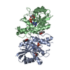
| Entry | Database: PDB / ID: 1rtw | ||||||
|---|---|---|---|---|---|---|---|
| Title | X-ray Structure of PF1337, a TenA Homologue from Pyrococcus furiosus. Northeast Structural Genomics Research Consortium (Nesg) Target PFR34 | ||||||
 Components Components | transcriptional activator, putative | ||||||
 Keywords Keywords | structural genomics / unknown function / pf1337 / tena / thiamin / PSI / Protein Structure Initiative / Northeast Structural Genomics Consortium / NESG | ||||||
| Function / homology |  Function and homology information Function and homology informationsmall molecule metabolic process / sulfur compound metabolic process / cytosol Similarity search - Function | ||||||
| Biological species |   Pyrococcus furiosus (archaea) Pyrococcus furiosus (archaea) | ||||||
| Method |  X-RAY DIFFRACTION / SYNCHROTRON / MAD / Resolution: 2.35 Å X-RAY DIFFRACTION / SYNCHROTRON / MAD / Resolution: 2.35 Å | ||||||
 Authors Authors | Benach, J. / Edstrom, W.C. / Lee, I. / Rong, X. / Acton, T.B. / Montelione, G.T. / Hunt, J.F. / Northeast Structural Genomics Consortium (NESG) | ||||||
 Citation Citation | Journal: Acta Crystallogr.,Sect.D / Year: 2005 Title: The 2.35 A structure of the TenA homolog from Pyrococcus furiosus supports an enzymatic function in thiamine metabolism. Authors: Benach, J. / Edstrom, W.C. / Lee, I. / Das, K. / Cooper, B. / Xiao, R. / Liu, J. / Rost, B. / Acton, T.B. / Montelione, G.T. / Hunt, J.F. | ||||||
| History |
| ||||||
| Remark 600 | ligand THE NATURE OF THE LIGAND IS TENTATIVE. THE LIGAND MP5 IS IS CLEARLY VISIBLE ONLY IN MONOMER ...ligand THE NATURE OF THE LIGAND IS TENTATIVE. THE LIGAND MP5 IS IS CLEARLY VISIBLE ONLY IN MONOMER A. IN THE OTHER MONOMERS, ONLY THE PHOSPHATE PART OF THE LIGAND HAS BEEN MODELLED. |
- Structure visualization
| Structure viewer | Molecule: MolmilJmol/JSmol |
|---|
- Downloads & links
-Download
| PDBx/mmCIF format | 1rtw.cif.gz | 186.3 KB | Display | PDBx/mmCIF format |
|---|---|---|---|---|
| PDB format | pdb1rtw.ent.gz | 152.2 KB | Display | PDB format |
| PDBx/mmJSON format | 1rtw.json.gz | Tree view | PDBx/mmJSON format | |
| Others |  Other downloads Other downloads |
-Validation report
| Summary document | 1rtw_validation.pdf.gz | 492.7 KB | Display | wwPDB validaton report |
|---|---|---|---|---|
| Full document | 1rtw_full_validation.pdf.gz | 514.2 KB | Display | |
| Data in XML | 1rtw_validation.xml.gz | 39.6 KB | Display | |
| Data in CIF | 1rtw_validation.cif.gz | 51.2 KB | Display | |
| Arichive directory | https://data.pdbj.org/pub/pdb/validation_reports/rt/1rtwftp://data.pdbj.org/pub/pdb/validation_reports/rt/1rtw | HTTPS FTP |
-Related structure data
| Similar structure data | |
|---|---|
| Other databases |


-Links
 PDBj
PDBj- Assembly
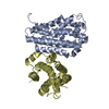
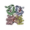
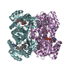
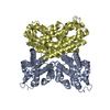
| Deposited unit | 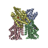
| ||||||||
|---|---|---|---|---|---|---|---|---|---|
| 1 |
| ||||||||
| Unit cell |
|
-Components
| #1: Protein | Mass: 26761.631 Da / Num. of mol.: 4 Source method: isolated from a genetically manipulated source Source: (gene. exp.) Pyrococcus furiosus (archaea) / Strain: DSM 3638 / Plasmid: pET21d / Production host:  Strain (production host): BL21(DE3) containing rare-tRNA expression plasmid pMGK References: UniProt: Q8U189 #2: Chemical | ChemComp-MP5 / ( | 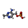  Mass: 219.135 Da / Num. of mol.: 1 / Source method: obtained synthetically / Formula: C6H10N3O4P Mass: 219.135 Da / Num. of mol.: 1 / Source method: obtained synthetically / Formula: C6H10N3O4P#3: Chemical |   Mass: 94.971 Da / Num. of mol.: 3 / Source method: obtained synthetically / Formula: PO4 Mass: 94.971 Da / Num. of mol.: 3 / Source method: obtained synthetically / Formula: PO4#4: Water | ChemComp-HOH / |  Mass: 18.015 Da / Num. of mol.: 234 / Source method: isolated from a natural source / Formula: H2O Mass: 18.015 Da / Num. of mol.: 234 / Source method: isolated from a natural source / Formula: H2OHas protein modification | Y | |
|---|
-Experimental details
-Experiment
| Experiment | Method: X-RAY DIFFRACTION / Number of used crystals: 1 |
|---|
- Sample preparation
| Crystal | Density Matthews: 2.4 Å3/Da / Density % sol: 48.67 % | |||||||||||||||||||||||||||||||||||||||||||||||||||||||||||||||
|---|---|---|---|---|---|---|---|---|---|---|---|---|---|---|---|---|---|---|---|---|---|---|---|---|---|---|---|---|---|---|---|---|---|---|---|---|---|---|---|---|---|---|---|---|---|---|---|---|---|---|---|---|---|---|---|---|---|---|---|---|---|---|---|---|
| Crystal grow | Temperature: 277 K / Method: vapor diffusion, hanging drop / pH: 7.5 Details: 20% PEG400, 200mM CaCl2, 50mM cacodylic acid, 1mM DTT, pH 7.5, VAPOR DIFFUSION, HANGING DROP, temperature 277K | |||||||||||||||||||||||||||||||||||||||||||||||||||||||||||||||
| Crystal grow | *PLUS Temperature: 277 K / pH: 8 / Method: vapor diffusion, hanging drop | |||||||||||||||||||||||||||||||||||||||||||||||||||||||||||||||
| Components of the solutions | *PLUS
|
-Data collection
| Diffraction | Mean temperature: 100 K | ||||||||||||
|---|---|---|---|---|---|---|---|---|---|---|---|---|---|
| Diffraction source | Source: SYNCHROTRON / Site: NSLS  / Beamline: X12C / Wavelength: 0.979, 0.9794, 0.95 / Beamline: X12C / Wavelength: 0.979, 0.9794, 0.95 | ||||||||||||
| Detector | Type: BRANDEIS - B4 / Detector: CCD / Date: Sep 12, 2003 | ||||||||||||
| Radiation | Monochromator: Si 111 / Protocol: MAD / Monochromatic (M) / Laue (L): M / Scattering type: x-ray | ||||||||||||
| Radiation wavelength |
| ||||||||||||
| Reflection | Resolution: 2.35→100 Å / Num. obs: 40075 / Observed criterion σ(F): -3 / Observed criterion σ(I): -3 / Redundancy: 3.7 % / Rmerge(I) obs: 0.071 / Net I/σ(I): 18.7 | ||||||||||||
| Reflection shell | Resolution: 2.35→2.38 Å / Rmerge(I) obs: 0.377 / Mean I/σ(I) obs: 3.7 | ||||||||||||
| Reflection | *PLUS Lowest resolution: 20 Å / Num. obs: 73637 / % possible obs: 93.5 % / Redundancy: 3.3 % / Num. measured all: 239958 / Rmerge(I) obs: 0.07 | ||||||||||||
| Reflection shell | *PLUS Lowest resolution: 2.43 Å / % possible obs: 89.7 % / Redundancy: 3.3 % / Num. unique obs: 6861 / Num. measured obs: 22349 / Rmerge(I) obs: 0.33 |
- Processing
| Software |
| ||||||||||||
|---|---|---|---|---|---|---|---|---|---|---|---|---|---|
| Refinement | Method to determine structure: MAD / Resolution: 2.35→20 Å / σ(F): 1 / Stereochemistry target values: Engh & Huber
| ||||||||||||
| Refinement step | Cycle: LAST / Resolution: 2.35→20 Å
| ||||||||||||
| Refinement | *PLUS Lowest resolution: 20 Å / Rfactor Rfree: 0.28 | ||||||||||||
| Solvent computation | *PLUS | ||||||||||||
| Displacement parameters | *PLUS | ||||||||||||
| Refine LS restraints | *PLUS
| ||||||||||||
| LS refinement shell | *PLUS Highest resolution: 2.35 Å / Lowest resolution: 2.43 Å / Rfactor Rfree: 0.31 / Rfactor Rwork: 0.27 |
