 Movie
Movie Controller

[English] 日本語
 Yorodumi
- PDB-1rru: The influence of a chiral amino acid on the helical handedness of... -
+ Open data

- Basic information
| Entry | Database: PDB / ID: 1rru | ||||||||||||||||||||||||
|---|---|---|---|---|---|---|---|---|---|---|---|---|---|---|---|---|---|---|---|---|---|---|---|---|---|
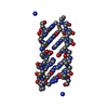
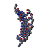
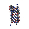
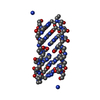
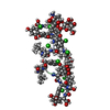
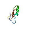
| Title | The influence of a chiral amino acid on the helical handedness of PNA in solution and in crystals | ||||||||||||||||||||||||
 Components Components | Peptide Nucleic Acid, (H-P(* Keywords KeywordsPEPTIDE NUCLEIC ACID / PNA / L-Lysine / helical handedness / P-form / molecular mechanics | Function / homology | PEP_NUC |  Function and homology information Function and homology informationMethod |  X-RAY DIFFRACTION / MOLECULAR REPLACEMENT / Resolution: 2.35 Å X-RAY DIFFRACTION / MOLECULAR REPLACEMENT / Resolution: 2.35 Å  Authors AuthorsRasmussen, H. / Liljefors, T. / Petersson, B. / Nielsen, P.E. / Kastrup, J.S. |  CitationJournal: J.Biomol.Struct.Dyn. / Year: 2004 CitationJournal: J.Biomol.Struct.Dyn. / Year: 2004Title: The Influence of a Chiral Amino Acid on the Helical Handedness of PNA in Solution and in Crystals Authors: Rasmussen, H. / Liljefors, T. / Petersson, B. / Nielsen, P.E. / Kastrup, J.S. #1: Journal: Nat.Struct.Biol. / Year: 1997Title: Crystal structure of a peptide nucleic acid (PNA) duplex at 1.7 A resolution Authors: Rasmussen, H. / Kastrup, J.S. / Nielsen, J.N. / Nielsen, J.M. / Nielsen, P.E. #2: Journal: New J.Chem. / Year: 1999Title: Peptide nucleic acids (PNA) derived from N-(N-methylaminoethyl) glycine. Synthesis, hybridization and structural properties Authors: Haiima, G. / Rasmussen, H. / Schmidt, G. / Jensen, D.K. / Kastrup, J.S. / Stafshede, P.W. / Norden, B. / Buckhardt, O. / Nielsen, P.E. #3: Journal: Eur.J.Org.Chem. / Year: 2001Title: 1,8-Naphthyridin-2(1H)-ones. Novel bi- and tricyclic analogues of Thymine in Peptide nucleic acids (PNA) Authors: Eldrup, A.B. / Nielsen, B.B. / Haiima, G. / Rasmussen, H. / Kastrup, J.S. / Christensen, C. / Nielsen, P.E. History |
|
- Structure visualization
| Structure viewer | Molecule: MolmilJmol/JSmol |
|---|
- Downloads & links
-Download
| PDBx/mmCIF format | 1rru.cif.gz | 19.2 KB | Display | PDBx/mmCIF format |
|---|---|---|---|---|
| PDB format | pdb1rru.ent.gz | 12.4 KB | Display | PDB format |
| PDBx/mmJSON format | 1rru.json.gz | Tree view | PDBx/mmJSON format | |
| Others |  Other downloads Other downloads |
-Validation report
| Arichive directory | https://data.pdbj.org/pub/pdb/validation_reports/rr/1rruftp://data.pdbj.org/pub/pdb/validation_reports/rr/1rru | HTTPS FTP |
|---|
-Related structure data
| Related structure data |  1pupS S: Starting model for refinement |
|---|---|
| Similar structure data |


-Links
 PDBj
PDBj- Assembly
| Deposited unit | 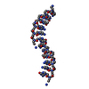
| ||||||||
|---|---|---|---|---|---|---|---|---|---|
| 1 | 
| ||||||||
| 2 | 
| ||||||||
| Unit cell |
|
-Components
| #1: Peptide nucleic acid | Mass: 1777.785 Da / Num. of mol.: 2 / Source method: obtained synthetically #2: Water | ChemComp-HOH / |  Mass: 18.015 Da / Num. of mol.: 28 / Source method: isolated from a natural source / Formula: H2O Mass: 18.015 Da / Num. of mol.: 28 / Source method: isolated from a natural source / Formula: H2OHas protein modification | Y | |
|---|
-Experimental details
-Experiment
| Experiment | Method: X-RAY DIFFRACTION / Number of used crystals: 1 |
|---|
- Sample preparation
| Crystal | Density Matthews: 3.68 Å3/Da / Density % sol: 66.62 % | ||||||||||||||||||||||||
|---|---|---|---|---|---|---|---|---|---|---|---|---|---|---|---|---|---|---|---|---|---|---|---|---|---|
| Crystal grow | Temperature: 293 K / Method: vapor diffusion, hanging drop Details: glycerol, magnesium formate, VAPOR DIFFUSION, HANGING DROP, temperature 293K | ||||||||||||||||||||||||
| Components of the solutions |
|
-Data collection
| Diffraction | Mean temperature: 293 K |
|---|---|
| Diffraction source | Source: ROTATING ANODE / Type: RIGAKU RU200 / Wavelength: 1.5418 Å |
| Detector | Type: RIGAKU RAXIS II / Detector: IMAGE PLATE / Date: Apr 16, 1997 |
| Radiation | Protocol: SINGLE WAVELENGTH / Monochromatic (M) / Laue (L): M / Scattering type: x-ray |
| Radiation wavelength | Wavelength: 1.5418 Å / Relative weight: 1 |
| Reflection | Resolution: 2.35→25 Å / Num. all: 2069 / Num. obs: 2069 / % possible obs: 98.8 % / Observed criterion σ(F): 0 / Observed criterion σ(I): 0 / Redundancy: 3.4 % / Rmerge(I) obs: 0.06 / Net I/σ(I): 15.9 |
| Reflection shell | Resolution: 2.35→2.39 Å / Redundancy: 3 % / Rmerge(I) obs: 0.316 / Mean I/σ(I) obs: 3.2 / Num. unique all: 92 / % possible all: 95.8 |
- Processing
| Software |
| |||||||||||||||||||||||||
|---|---|---|---|---|---|---|---|---|---|---|---|---|---|---|---|---|---|---|---|---|---|---|---|---|---|---|
| Refinement | Method to determine structure: MOLECULAR REPLACEMENT Starting model: PDB entry 1PUP Resolution: 2.35→6 Å / Isotropic thermal model: isotropic / Cross valid method: troughout / σ(F): 3 / Stereochemistry target values: Engh & Huber / Details: Own parameter and topology file created
| |||||||||||||||||||||||||
| Displacement parameters | Biso mean: 38.4 Å2 | |||||||||||||||||||||||||
| Refinement step | Cycle: LAST / Resolution: 2.35→6 Å
| |||||||||||||||||||||||||
| Refine LS restraints |
| |||||||||||||||||||||||||
| LS refinement shell | Resolution: 2.35→2.45 Å /
|
