 Movie
Movie Controller

[English] 日本語
 Yorodumi
- PDB-1rqy: 9-amino-[N-(2-dimethylamino)proply]-acridine-4-carboxamide bound ... -
+ Open data

- Basic information
| Entry | Database: PDB / ID: 1rqy | ||||||||||||||||||
|---|---|---|---|---|---|---|---|---|---|---|---|---|---|---|---|---|---|---|---|
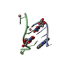
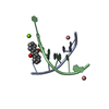
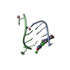
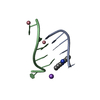
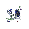
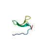
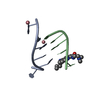
| Title | 9-amino-[N-(2-dimethylamino)proply]-acridine-4-carboxamide bound to d(CGTACG)2 | ||||||||||||||||||
 Components Components | 5'-D(CP* Keywords KeywordsDNA / B-DNA / cytosine exchange / quadruplex / acridine / unusual intercalation | Function / homology | Chem-7AD / : / DNA |  Function and homology information Function and homology informationMethod |  X-RAY DIFFRACTION / refinement / Resolution: 1.55 Å X-RAY DIFFRACTION / refinement / Resolution: 1.55 Å  Authors AuthorsAdams, A. / Guss, J.M. / Denny, W.A. / Wakelin, L.P. |  CitationJournal: Acta Crystallogr.,Sect.D / Year: 2004 CitationJournal: Acta Crystallogr.,Sect.D / Year: 2004Title: Structure of 9-amino-[N-(2-dimethylamino)propyl]acridine-4-carboxamide bound to d(CGTACG)(2): a comparison of structures of d(CGTACG)(2) complexed with intercalatorsin the presence of cobalt. Authors: Adams, A. / Guss, J.M. / Denny, W.A. / Wakelin, L.P. History |
|
- Structure visualization
| Structure viewer | Molecule: MolmilJmol/JSmol |
|---|
- Downloads & links
-Download
| PDBx/mmCIF format | 1rqy.cif.gz | 20.7 KB | Display | PDBx/mmCIF format |
|---|---|---|---|---|
| PDB format | pdb1rqy.ent.gz | 11.9 KB | Display | PDB format |
| PDBx/mmJSON format | 1rqy.json.gz | Tree view | PDBx/mmJSON format | |
| Others |  Other downloads Other downloads |
-Validation report
| Arichive directory | https://data.pdbj.org/pub/pdb/validation_reports/rq/1rqyftp://data.pdbj.org/pub/pdb/validation_reports/rq/1rqy | HTTPS FTP |
|---|
-Related structure data
| Related structure data |  1fn1S S: Starting model for refinement |
|---|---|
| Similar structure data |


-Links
 PDBj
PDBj


- Assembly
| Deposited unit | 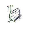
| ||||||||||||||||||
|---|---|---|---|---|---|---|---|---|---|---|---|---|---|---|---|---|---|---|---|
| 1 |
| ||||||||||||||||||
| Unit cell |
| ||||||||||||||||||
| Components on special symmetry positions |
|
-Components
| #1: DNA chain | Mass: 1809.217 Da / Num. of mol.: 2 / Source method: obtained synthetically #2: Chemical |   Mass: 58.933 Da / Num. of mol.: 2 / Source method: obtained synthetically / Formula: Co Mass: 58.933 Da / Num. of mol.: 2 / Source method: obtained synthetically / Formula: Co#3: Chemical |   Mass: 322.404 Da / Num. of mol.: 3 / Source method: obtained synthetically / Formula: C19H22N4O Mass: 322.404 Da / Num. of mol.: 3 / Source method: obtained synthetically / Formula: C19H22N4O#4: Chemical |   Mass: 24.305 Da / Num. of mol.: 2 / Source method: obtained synthetically / Formula: Mg Mass: 24.305 Da / Num. of mol.: 2 / Source method: obtained synthetically / Formula: Mg#5: Water | ChemComp-HOH / |  Mass: 18.015 Da / Num. of mol.: 30 / Source method: isolated from a natural source / Formula: H2O Mass: 18.015 Da / Num. of mol.: 30 / Source method: isolated from a natural source / Formula: H2O |
|---|
-Experimental details
-Experiment
| Experiment | Method: X-RAY DIFFRACTION / Number of used crystals: 1 |
|---|
- Sample preparation
| Crystal | Density Matthews: 2 Å3/Da / Density % sol: 37.91 % | ||||||||||||||||||||||||||||||||||||||||||||||||
|---|---|---|---|---|---|---|---|---|---|---|---|---|---|---|---|---|---|---|---|---|---|---|---|---|---|---|---|---|---|---|---|---|---|---|---|---|---|---|---|---|---|---|---|---|---|---|---|---|---|
| Crystal grow | Temperature: 285 K / Method: vapor diffusion, sitting drop / pH: 6.5 Details: sodium cacodylate, magnesium acetate, cobalt chloride, spermine, MPD, pH 6.5, VAPOR DIFFUSION, SITTING DROP, temperature 285K | ||||||||||||||||||||||||||||||||||||||||||||||||
| Components of the solutions |
|
-Data collection
| Diffraction | Mean temperature: 110 K |
|---|---|
| Diffraction source | Source: ROTATING ANODE / Type: RIGAKU RU200 / Wavelength: 1.5418 Å |
| Detector | Type: MARRESEARCH / Detector: AREA DETECTOR / Date: Mar 18, 1998 / Details: focusing mirror optics |
| Radiation | Monochromator: Osmic mirrors / Protocol: SINGLE WAVELENGTH / Monochromatic (M) / Laue (L): M / Scattering type: x-ray |
| Radiation wavelength | Wavelength: 1.5418 Å / Relative weight: 1 |
| Reflection | Resolution: 1.55→40 Å / Num. all: 4786 / Num. obs: 4786 / % possible obs: 97.6 % / Observed criterion σ(F): 0 / Observed criterion σ(I): 0 / Redundancy: 9.8 % / Biso Wilson estimate: 20.6 Å2 / Rsym value: 0.077 / Net I/σ(I): 12 |
| Reflection shell | Resolution: 1.55→1.61 Å / Redundancy: 3.8 % / Mean I/σ(I) obs: 3.8 / Num. unique all: 383 / Rsym value: 0.26 / % possible all: 81.7 |
- Processing
| Software |
| |||||||||||||||||||||||||||||||||
|---|---|---|---|---|---|---|---|---|---|---|---|---|---|---|---|---|---|---|---|---|---|---|---|---|---|---|---|---|---|---|---|---|---|---|
| Refinement | Method to determine structure: refinement Starting model: PDB entry 1FN1 Resolution: 1.55→40 Å / Num. parameters: 1235 / Num. restraintsaints: 2833 / Cross valid method: FREE R / σ(F): 0 / σ(I): 0 / Stereochemistry target values: ENGH AND HUBER Details: ANISOTROPIC SCALING APPLIED BY THE METHOD OF PARKIN, MOEZZI & HOPE, J.APPL.CRYST.28(1995)53-56
| |||||||||||||||||||||||||||||||||
| Refine analyze | Num. disordered residues: 3 / Occupancy sum hydrogen: 0 / Occupancy sum non hydrogen: 269.25 | |||||||||||||||||||||||||||||||||
| Refinement step | Cycle: LAST / Resolution: 1.55→40 Å
| |||||||||||||||||||||||||||||||||
| Refine LS restraints |
| |||||||||||||||||||||||||||||||||
| LS refinement shell | Resolution: 1.55→1.6 Å /
|
