 Movie
Movie Controller

[English] 日本語
 Yorodumi
- PDB-1rpj: CRYSTAL STRUCTURE OF D-ALLOSE BINDING PROTEIN FROM ESCHERICHIA COLI -
+ Open data

- Basic information
| Entry | Database: PDB / ID: 1rpj | |||||||||
|---|---|---|---|---|---|---|---|---|---|---|
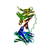
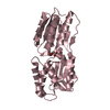
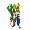
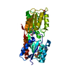
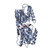
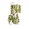
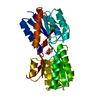
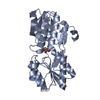
| Title | CRYSTAL STRUCTURE OF D-ALLOSE BINDING PROTEIN FROM ESCHERICHIA COLI | |||||||||
 Components Components | PROTEIN (PRECURSOR OF PERIPLASMIC SUGAR RECEPTOR) | |||||||||
 Keywords Keywords | PERIPLASMIC SUGAR RECEPTOR | |||||||||
| Function / homology |  Function and homology information Function and homology informationD-allose transmembrane transport / D-ribose transmembrane transport / monosaccharide binding / ATP-binding cassette (ABC) transporter complex, substrate-binding subunit-containing / transmembrane transport / outer membrane-bounded periplasmic space / membrane Similarity search - Function | |||||||||
| Biological species |  | |||||||||
| Method |  X-RAY DIFFRACTION / MOLECULAR REPLACEMENT / Resolution: 1.8 Å X-RAY DIFFRACTION / MOLECULAR REPLACEMENT / Resolution: 1.8 Å | |||||||||
 Authors Authors | Chaudhuri, B. / Jones, T.A. / Mowbray, S.L. | |||||||||
 Citation Citation | Journal: J.Mol.Biol. / Year: 1999 Title: Structure of D-allose binding protein from Escherichia coli bound to D-allose at 1.8 A resolution. Authors: Chaudhuri, B.N. / Ko, J. / Park, C. / Jones, T.A. / Mowbray, S.L. #1: Journal: J.Bacteriol. / Year: 1997Title: The D-Allose Operon of Escherichia Coli K-12 Authors: Kim, C. / Song, S. / Park, C. | |||||||||
| History |
|
- Structure visualization
| Structure viewer | Molecule: MolmilJmol/JSmol |
|---|
- Downloads & links
-Download
| PDBx/mmCIF format | 1rpj.cif.gz | 72.1 KB | Display | PDBx/mmCIF format |
|---|---|---|---|---|
| PDB format | pdb1rpj.ent.gz | 51.2 KB | Display | PDB format |
| PDBx/mmJSON format | 1rpj.json.gz | Tree view | PDBx/mmJSON format | |
| Others |  Other downloads Other downloads |
-Validation report
| Summary document | 1rpj_validation.pdf.gz | 444.5 KB | Display | wwPDB validaton report |
|---|---|---|---|---|
| Full document | 1rpj_full_validation.pdf.gz | 446.7 KB | Display | |
| Data in XML | 1rpj_validation.xml.gz | 14.4 KB | Display | |
| Data in CIF | 1rpj_validation.cif.gz | 20.9 KB | Display | |
| Arichive directory | https://data.pdbj.org/pub/pdb/validation_reports/rp/1rpjftp://data.pdbj.org/pub/pdb/validation_reports/rp/1rpj | HTTPS FTP |
-Related structure data
| Related structure data |  1gubC  1gudC  2driS S: Starting model for refinement C: citing same article ( |
|---|---|
| Similar structure data |


-Links
 PDBj
PDBj- Assembly
| Deposited unit | 
| ||||||||||
|---|---|---|---|---|---|---|---|---|---|---|---|
| 1 |
| ||||||||||
| Unit cell |
|
-Components
| #1: Protein | Mass: 30417.807 Da / Num. of mol.: 1 / Source method: isolated from a natural source / Source: (natural) |
|---|---|
| #2: Chemical | ChemComp-ZN /   Mass: 65.409 Da / Num. of mol.: 1 / Source method: obtained synthetically / Formula: Zn Mass: 65.409 Da / Num. of mol.: 1 / Source method: obtained synthetically / Formula: Zn |
| #3: Chemical | ChemComp-SO4 /   Mass: 96.063 Da / Num. of mol.: 1 / Source method: obtained synthetically / Formula: SO4 Mass: 96.063 Da / Num. of mol.: 1 / Source method: obtained synthetically / Formula: SO4 |
| #4: Sugar | ChemComp-ALL /   Type: D-saccharide, beta linking / Mass: 180.156 Da / Num. of mol.: 1 / Source method: obtained synthetically / Formula: C6H12O6 Type: D-saccharide, beta linking / Mass: 180.156 Da / Num. of mol.: 1 / Source method: obtained synthetically / Formula: C6H12O6 |
| #5: Water | ChemComp-HOH /  Mass: 18.015 Da / Num. of mol.: 215 / Source method: isolated from a natural source / Formula: H2O Mass: 18.015 Da / Num. of mol.: 215 / Source method: isolated from a natural source / Formula: H2O |
| Nonpolymer details | ZINC SULFATE WAS ESSENTIAL FOR CRYSTALLIZATION AND BOTH ZINC AND SULFATE IONS COULD BE MODELLED IN ...ZINC SULFATE WAS ESSENTIAL FOR CRYSTALLIZ |
-Experimental details
-Experiment
| Experiment | Method: X-RAY DIFFRACTION / Number of used crystals: 1 |
|---|
- Sample preparation
| Crystal | Density Matthews: 1.98 Å3/Da / Density % sol: 41 % | ||||||||||||||||||||||||||||||
|---|---|---|---|---|---|---|---|---|---|---|---|---|---|---|---|---|---|---|---|---|---|---|---|---|---|---|---|---|---|---|---|
| Crystal grow | pH: 6.5 / Details: pH 6.5 | ||||||||||||||||||||||||||||||
| Components of the solutions |
| ||||||||||||||||||||||||||||||
| Crystal grow | *PLUS Method: vapor diffusion, hanging drop | ||||||||||||||||||||||||||||||
| Components of the solutions | *PLUS
|
-Data collection
| Diffraction | Mean temperature: 100 K |
|---|---|
| Diffraction source | Source: ROTATING ANODE / Type: RIGAKU / Wavelength: 1.5418 |
| Detector | Type: RIGAKU RAXIS IIC / Detector: IMAGE PLATE |
| Radiation | Monochromator: GRAPHITE / Protocol: SINGLE WAVELENGTH / Monochromatic (M) / Laue (L): M / Scattering type: x-ray |
| Radiation wavelength | Wavelength: 1.5418 Å / Relative weight: 1 |
| Reflection | Resolution: 1.8→29.787 Å / Num. obs: 21163 / % possible obs: 96.5 % / Redundancy: 2.8 % / Biso Wilson estimate: 17.07 Å2 / Rmerge(I) obs: 0.045 / Net I/σ(I): 10.4 |
| Reflection shell | Resolution: 1.8→1.85 Å / Redundancy: 2.4 % / Rmerge(I) obs: 0.15 / Mean I/σ(I) obs: 5 / % possible all: 93.6 |
| Reflection shell | *PLUS % possible obs: 93.6 % |
- Processing
| Software |
| ||||||||||||||||||||||||||||||||||||||||||||||||||||||||||||||||||||||||||||||||||||
|---|---|---|---|---|---|---|---|---|---|---|---|---|---|---|---|---|---|---|---|---|---|---|---|---|---|---|---|---|---|---|---|---|---|---|---|---|---|---|---|---|---|---|---|---|---|---|---|---|---|---|---|---|---|---|---|---|---|---|---|---|---|---|---|---|---|---|---|---|---|---|---|---|---|---|---|---|---|---|---|---|---|---|---|---|---|
| Refinement | Method to determine structure: MOLECULAR REPLACEMENT Starting model: PDB ENTRY 2DRI Resolution: 1.8→29 Å / Cross valid method: THROUGHOUT / σ(F): 0 / Details: CNS WAS USED BEFORE REFMAC FOR REFINEMENT
| ||||||||||||||||||||||||||||||||||||||||||||||||||||||||||||||||||||||||||||||||||||
| Displacement parameters | Biso mean: 11.18 Å2 | ||||||||||||||||||||||||||||||||||||||||||||||||||||||||||||||||||||||||||||||||||||
| Refinement step | Cycle: LAST / Resolution: 1.8→29 Å
| ||||||||||||||||||||||||||||||||||||||||||||||||||||||||||||||||||||||||||||||||||||
| Refine LS restraints |
| ||||||||||||||||||||||||||||||||||||||||||||||||||||||||||||||||||||||||||||||||||||
| Software | *PLUS Name: REFMAC / Classification: refinement | ||||||||||||||||||||||||||||||||||||||||||||||||||||||||||||||||||||||||||||||||||||
| Refinement | *PLUS Rfactor obs: 0.194 | ||||||||||||||||||||||||||||||||||||||||||||||||||||||||||||||||||||||||||||||||||||
| Solvent computation | *PLUS | ||||||||||||||||||||||||||||||||||||||||||||||||||||||||||||||||||||||||||||||||||||
| Displacement parameters | *PLUS |
