 Movie
Movie Controller

[English] 日本語
 Yorodumi
- PDB-1rjr: The crystal structure of the D-aminoacylase D366A mutant in compl... -
+ Open data

- Basic information
| Entry | Database: PDB / ID: 1rjr | ||||||
|---|---|---|---|---|---|---|---|
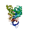
| Title | The crystal structure of the D-aminoacylase D366A mutant in complex with 100mM ZnCl2 | ||||||
 Components Components | D-aminoacylase | ||||||
 Keywords Keywords | HYDROLASE / TIM barrel / beta barrel / insertion | ||||||
| Function / homology |  Function and homology information Function and homology informationN-acyl-D-amino-acid deacylase / N-acyl-D-amino-acid deacylase activity / hydrolase activity, acting on carbon-nitrogen (but not peptide) bonds, in cyclic amides / metal ion binding / cytosol Similarity search - Function | ||||||
| Biological species |  Alcaligenes faecalis (bacteria) Alcaligenes faecalis (bacteria) | ||||||
| Method |  X-RAY DIFFRACTION / SYNCHROTRON / MOLECULAR REPLACEMENT / Resolution: 2.1 Å X-RAY DIFFRACTION / SYNCHROTRON / MOLECULAR REPLACEMENT / Resolution: 2.1 Å | ||||||
 Authors Authors | Lai, W.L. / Chou, L.Y. / Ting, C.Y. / Tsai, Y.C. / Liaw, S.H. | ||||||
 Citation Citation | Journal: J.Biol.Chem. / Year: 2004 Title: The functional role of the binuclear metal center in D-aminoacylase: one-metal activation and second-metal attenuation. Authors: Lai, W.L. / Chou, L.Y. / Ting, C.Y. / Kirby, R. / Tsai, Y.C. / Wang, A.H. / Liaw, S.H. #1: Journal: J.Biol.Chem. / Year: 2003Title: Crystal structure of D-aminoacylase from Alcaligenes faecalis DA1. A novel subset of amidohydrolases and insights into the enzyme mechanism Authors: Liaw, S.H. / Chen, S.J. / Ko, T.P. / Hsu, C.S. / Chen, C.J. / Wang, A.H. / Tsai, Y.C. #2: Journal: Protein Sci. / Year: 2002 Title: Structural-based mutational analysis of D-aminoacylase from Alcaligenes faecalis DA1 Authors: Hsu, C.S. / Lai, W.L. / Chang, W.W. / Liaw, S.H. / Tsai, Y.C. | ||||||
| History |
|
- Structure visualization
| Structure viewer | Molecule: MolmilJmol/JSmol |
|---|
- Downloads & links
-Download
| PDBx/mmCIF format | 1rjr.cif.gz | 107.2 KB | Display | PDBx/mmCIF format |
|---|---|---|---|---|
| PDB format | pdb1rjr.ent.gz | 79.6 KB | Display | PDB format |
| PDBx/mmJSON format | 1rjr.json.gz | Tree view | PDBx/mmJSON format | |
| Others |  Other downloads Other downloads |
-Validation report
| Arichive directory | https://data.pdbj.org/pub/pdb/validation_reports/rj/1rjrftp://data.pdbj.org/pub/pdb/validation_reports/rj/1rjr | HTTPS FTP |
|---|
-Related structure data
| Related structure data |  1rjpC  1rjqC 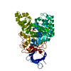 1rk5C 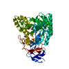 1rk6C 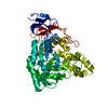 1v4yC  1v51C  1m7jS S: Starting model for refinement C: citing same article ( |
|---|---|
| Similar structure data |


-Links
 PDBj
PDBj

- Assembly
| Deposited unit | 
| ||||||||
|---|---|---|---|---|---|---|---|---|---|
| 1 |
| ||||||||
| Unit cell |
|
-Components
| #1: Protein | Mass: 53422.227 Da / Num. of mol.: 1 / Mutation: D366A Source method: isolated from a genetically manipulated source Source: (gene. exp.) Alcaligenes faecalis (bacteria) / Gene: DA1 / Plasmid: pQE30 / Production host: | ||||
|---|---|---|---|---|---|
| #2: Chemical |   Mass: 59.044 Da / Num. of mol.: 2 / Source method: obtained synthetically / Formula: C2H3O2 Mass: 59.044 Da / Num. of mol.: 2 / Source method: obtained synthetically / Formula: C2H3O2#3: Chemical |   Mass: 65.409 Da / Num. of mol.: 2 / Source method: obtained synthetically / Formula: Zn Mass: 65.409 Da / Num. of mol.: 2 / Source method: obtained synthetically / Formula: Zn#4: Water | ChemComp-HOH / |  Mass: 18.015 Da / Num. of mol.: 176 / Source method: isolated from a natural source / Formula: H2O Mass: 18.015 Da / Num. of mol.: 176 / Source method: isolated from a natural source / Formula: H2O |
-Experimental details
-Experiment
| Experiment | Method: X-RAY DIFFRACTION / Number of used crystals: 1 |
|---|
- Sample preparation
| Crystal | Density Matthews: 3.2 Å3/Da / Density % sol: 61.21 % |
|---|---|
| Crystal grow | Temperature: 295 K / Method: vapor diffusion, hanging drop / pH: 5.6 Details: PEG4000, ammonium acetate, sodium citrate, pH 5.6, VAPOR DIFFUSION, HANGING DROP, temperature 295K |
-Data collection
| Diffraction | Mean temperature: 100 K |
|---|---|
| Diffraction source | Source: SYNCHROTRON / Site: NSRRC  / Beamline: BL17B2 / Wavelength: 1 Å / Beamline: BL17B2 / Wavelength: 1 Å |
| Detector | Type: RIGAKU RAXIS IV / Detector: IMAGE PLATE / Date: Apr 5, 2003 / Details: mirror |
| Radiation | Protocol: SINGLE WAVELENGTH / Monochromatic (M) / Laue (L): M / Scattering type: x-ray |
| Radiation wavelength | Wavelength: 1 Å / Relative weight: 1 |
| Reflection | Resolution: 2.1→50 Å / Num. all: 87018 / Num. obs: 34438 / % possible obs: 90.7 % / Observed criterion σ(F): 0 / Observed criterion σ(I): 0 / Redundancy: 2.5 % / Rmerge(I) obs: 0.044 / Rsym value: 0.04 / Net I/σ(I): 19.6 |
| Reflection shell | Resolution: 2.1→2.18 Å / Redundancy: 2.1 % / Rmerge(I) obs: 0.19 / Mean I/σ(I) obs: 4.3 / Num. unique all: 3228 / Rsym value: 0.198 / % possible all: 86 |
- Processing
| Software |
| |||||||||||||||||||||||||
|---|---|---|---|---|---|---|---|---|---|---|---|---|---|---|---|---|---|---|---|---|---|---|---|---|---|---|
| Refinement | Method to determine structure: MOLECULAR REPLACEMENT Starting model: 1M7J Resolution: 2.1→50 Å / Isotropic thermal model: isotropic B / Cross valid method: THROUGHOUT / σ(F): 0 / σ(I): 0 / Stereochemistry target values: Engh & Huber
| |||||||||||||||||||||||||
| Displacement parameters | Biso mean: 23 Å2 | |||||||||||||||||||||||||
| Refinement step | Cycle: LAST / Resolution: 2.1→50 Å
| |||||||||||||||||||||||||
| Refine LS restraints |
| |||||||||||||||||||||||||
| LS refinement shell | Resolution: 2.1→2.18 Å
|
