 Movie
Movie Controller

[English] 日本語
 Yorodumi
- PDB-1r9w: Crystal Structure of the DNA-binding domain of the human papillom... -
+ Open data

- Basic information
| Entry | Database: PDB / ID: 1r9w | ||||||
|---|---|---|---|---|---|---|---|
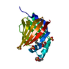
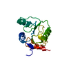
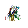
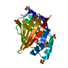
| Title | Crystal Structure of the DNA-binding domain of the human papillomavirus type 18 (HPV-18) replication initiation protein E1 | ||||||
 Components Components | Replication protein E1 | ||||||
 Keywords Keywords | REPLICATION / HPV-18 / papillomavirus / DNA-binding domain / viral replication / initiator protein | ||||||
| Function / homology |  Function and homology information Function and homology information3'-5' DNA helicase activity / DNA 3'-5' helicase / viral genome replication / DNA helicase activity / host cell cytoplasm / DNA replication / host cell nucleus / ATP hydrolysis activity / DNA binding / ATP binding Similarity search - Function | ||||||
| Biological species |  Human papillomavirus type 18 Human papillomavirus type 18 | ||||||
| Method |  X-RAY DIFFRACTION / SYNCHROTRON / MOLECULAR REPLACEMENT / Resolution: 1.8 Å X-RAY DIFFRACTION / SYNCHROTRON / MOLECULAR REPLACEMENT / Resolution: 1.8 Å | ||||||
 Authors Authors | Auster, A.S. / Joshua-Tor, L. | ||||||
 Citation Citation | Journal: J.Biol.Chem. / Year: 2004 Title: The DNA-binding domain of human papillomavirus type 18 E1. Crystal structure, dimerization, and DNA binding. Authors: Auster, A.S. / Joshua-Tor, L. | ||||||
| History |
|
- Structure visualization
| Structure viewer | Molecule: MolmilJmol/JSmol |
|---|
- Downloads & links
-Download
| PDBx/mmCIF format | 1r9w.cif.gz | 42.7 KB | Display | PDBx/mmCIF format |
|---|---|---|---|---|
| PDB format | pdb1r9w.ent.gz | 29 KB | Display | PDB format |
| PDBx/mmJSON format | 1r9w.json.gz | Tree view | PDBx/mmJSON format | |
| Others |  Other downloads Other downloads |
-Validation report
| Arichive directory | https://data.pdbj.org/pub/pdb/validation_reports/r9/1r9wftp://data.pdbj.org/pub/pdb/validation_reports/r9/1r9w | HTTPS FTP |
|---|
-Related structure data
| Related structure data |  1f08S S: Starting model for refinement |
|---|---|
| Similar structure data |


-Links
 PDBj
PDBj


- Assembly
| Deposited unit | 
| ||||||||
|---|---|---|---|---|---|---|---|---|---|
| 1 |
| ||||||||
| Unit cell |
| ||||||||
| Details | The biological assembly is a monomer. There is one monomer in the asymmetric unit. |
-Components
| #1: Protein | Mass: 16335.233 Da / Num. of mol.: 1 / Fragment: DNA-binding domain Source method: isolated from a genetically manipulated source Source: (gene. exp.) Human papillomavirus type 18 / Genus: Alphapapillomavirus / Species: Human papillomavirus - 18 / Gene: E1Plasmid details: constructed with a glutathione S-transferase (GST) cassette and a thrombin cleavage site Plasmid: pET11C / Species (production host): Escherichia coli / Production host:  |
|---|---|
| #2: Water | ChemComp-HOH /  Mass: 18.015 Da / Num. of mol.: 109 / Source method: isolated from a natural source / Formula: H2O Mass: 18.015 Da / Num. of mol.: 109 / Source method: isolated from a natural source / Formula: H2O |
-Experimental details
-Experiment
| Experiment | Method: X-RAY DIFFRACTION / Number of used crystals: 1 |
|---|
- Sample preparation
| Crystal | Density Matthews: 2.15 Å3/Da / Density % sol: 42.79 % | |||||||||||||||||||||||||||||||||||||||||||||||||||||||||||||||
|---|---|---|---|---|---|---|---|---|---|---|---|---|---|---|---|---|---|---|---|---|---|---|---|---|---|---|---|---|---|---|---|---|---|---|---|---|---|---|---|---|---|---|---|---|---|---|---|---|---|---|---|---|---|---|---|---|---|---|---|---|---|---|---|---|
| Crystal grow | Temperature: 290 K / Method: vapor diffusion, hanging drop / pH: 5 Details: PEG 4000, ammonium acetate, sodium citrate, DTT, pH 5.0, VAPOR DIFFUSION, HANGING DROP, temperature 290.0K | |||||||||||||||||||||||||||||||||||||||||||||||||||||||||||||||
| Crystal grow | *PLUS Temperature: 17 ℃ / pH: 7.5 / Method: vapor diffusion, hanging drop | |||||||||||||||||||||||||||||||||||||||||||||||||||||||||||||||
| Components of the solutions | *PLUS
|
-Data collection
| Diffraction | Mean temperature: 100 K |
|---|---|
| Diffraction source | Source: SYNCHROTRON / Site: NSLS  / Beamline: X26C / Wavelength: 1.1 Å / Beamline: X26C / Wavelength: 1.1 Å |
| Detector | Type: ADSC QUANTUM 4 / Detector: CCD / Details: mirrors |
| Radiation | Protocol: SINGLE WAVELENGTH / Monochromatic (M) / Laue (L): M / Scattering type: x-ray |
| Radiation wavelength | Wavelength: 1.1 Å / Relative weight: 1 |
| Reflection | Resolution: 1.8→30 Å / Num. obs: 12094 / % possible obs: 93.3 % / Observed criterion σ(I): -3 / Redundancy: 3.41 % / Biso Wilson estimate: 12.7 Å2 / Rmerge(I) obs: 0.049 / Net I/σ(I): 17.5 |
| Reflection shell | Resolution: 1.8→1.91 Å / Redundancy: 2.25 % / Rmerge(I) obs: 0.135 / Mean I/σ(I) obs: 5.7 / Num. unique all: 1412 / % possible all: 69.1 |
| Reflection | *PLUS % possible obs: 93 % / Num. measured all: 45127 |
| Reflection shell | *PLUS % possible obs: 69.1 % / Num. unique obs: 1412 |
- Processing
| Software |
| |||||||||||||||||||||||||
|---|---|---|---|---|---|---|---|---|---|---|---|---|---|---|---|---|---|---|---|---|---|---|---|---|---|---|
| Refinement | Method to determine structure: MOLECULAR REPLACEMENT Starting model: PDB entry 1F08 with non-identical residues mutated to alanines, and no solvent Resolution: 1.8→30 Å / Isotropic thermal model: isotropic / Cross valid method: THROUGHOUT / σ(F): 0 / Stereochemistry target values: Engh & Huber Details: Deposited structure factor file includes data to 1.5 A, however, statistics and refinement are reported to 1.8 A.
| |||||||||||||||||||||||||
| Displacement parameters | Biso mean: 19.4 Å2 | |||||||||||||||||||||||||
| Refine analyze |
| |||||||||||||||||||||||||
| Refinement step | Cycle: LAST / Resolution: 1.8→30 Å
| |||||||||||||||||||||||||
| Refine LS restraints |
| |||||||||||||||||||||||||
| LS refinement shell | Resolution: 1.8→1.91 Å / Rfactor Rfree error: 0.04
| |||||||||||||||||||||||||
| Refine LS restraints | *PLUS
|
