Resolution: 1.45→1.48 Å / Rmerge(I) obs: 0.468 / % possible all: 49
-
Processing
Software
Name
Classification
SHELX
modelbuilding
SHELXL-97
refinement
DENZO
datareduction
SCALEPACK
datascaling
SHELX
phasing
Refinement
Method to determine structure: AB INITIO PHASING Starting model: xylanase T-6 in hexagonal crystal form at 2.4A resolution Resolution: 1.45→40 Å / Num. parameters: 176 / Num. restraintsaints: 42293 / Cross valid method: FREE R / σ(F): 0 / Details: ANISOTROPIC REFINEMENT REDUCED FREE R (NO CUTOFF)
Rfactor
Num. reflection
% reflection
Selection details
Rfree
0.1808
3688
5.3 %
RANDOM
obs
0.1264
73607
94.1 %
-
all
-
95991
-
-
Refine analyze
Num. disordered residues: 34 / Occupancy sum hydrogen: 0 / Occupancy sum non hydrogen: 3616.09
Refinement step
Cycle: LAST / Resolution: 1.45→40 Å
Protein
Nucleic acid
Ligand
Solvent
Total
Num. atoms
3028
0
138
494
3660
Refine LS restraints
Refine-ID
Type
Dev ideal
X-RAY DIFFRACTION
s_bond_d
0.011
X-RAY DIFFRACTION
s_angle_d
0.029
X-RAY DIFFRACTION
s_similar_dist
0
X-RAY DIFFRACTION
s_from_restr_planes
0.028
X-RAY DIFFRACTION
s_zero_chiral_vol
0.055
X-RAY DIFFRACTION
s_non_zero_chiral_vol
0.06
X-RAY DIFFRACTION
s_anti_bump_dis_restr
0.015
X-RAY DIFFRACTION
s_rigid_bond_adp_cmpnt
0.003
X-RAY DIFFRACTION
s_similar_adp_cmpnt
0.035
X-RAY DIFFRACTION
s_approx_iso_adps
0.093
LS refinement shell
Resolution: 1.45→1.48 Å
+
About Yorodumi
-
News
-
Feb 9, 2022. New format data for meta-information of EMDB entries
New format data for meta-information of EMDB entries
Version 3 of the EMDB header file is now the official format.
The previous official version 1.9 will be removed from the archive.
In the structure databanks used in Yorodumi, some data are registered as the other names, "COVID-19 virus" and "2019-nCoV". Here are the details of the virus and the list of structure data.
Jan 31, 2019. EMDB accession codes are about to change! (news from PDBe EMDB page)
EMDB accession codes are about to change! (news from PDBe EMDB page)
The allocation of 4 digits for EMDB accession codes will soon come to an end. Whilst these codes will remain in use, new EMDB accession codes will include an additional digit and will expand incrementally as the available range of codes is exhausted. The current 4-digit format prefixed with “EMD-” (i.e. EMD-XXXX) will advance to a 5-digit format (i.e. EMD-XXXXX), and so on. It is currently estimated that the 4-digit codes will be depleted around Spring 2019, at which point the 5-digit format will come into force.
The EM Navigator/Yorodumi systems omit the EMD- prefix.
Related info.:Q: What is EMD? / ID/Accession-code notation in Yorodumi/EM Navigator
Yorodumi is a browser for structure data from EMDB, PDB, SASBDB, etc.
This page is also the successor to EM Navigator detail page, and also detail information page/front-end page for Omokage search.
The word "yorodu" (or yorozu) is an old Japanese word meaning "ten thousand". "mi" (miru) is to see.
Related info.:EMDB / PDB / SASBDB / Comparison of 3 databanks / Yorodumi Search / Aug 31, 2016. New EM Navigator & Yorodumi / Yorodumi Papers / Jmol/JSmol / Function and homology information / Changes in new EM Navigator and Yorodumi
 Movie
Movie Controller

 Yorodumi
 Open data
 Basic information
 Components
 Keywords
 Function and homology information
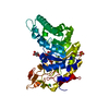
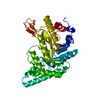
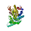
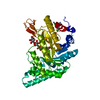
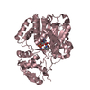
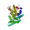
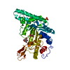
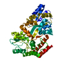
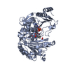
 Geobacillus stearothermophilus (bacteria)
 X-RAY DIFFRACTION /
  Authors
 Citation
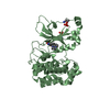
 Structure visualization
 Downloads & links
 Other downloads

 PDBj
PDBj
 Assembly
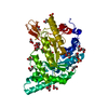


 Mass: 65.409 Da / Num. of mol.: 7 / Source method: obtained synthetically / Formula: Zn
 Mass: 35.453 Da / Num. of mol.: 2 / Source method: obtained synthetically / Formula: Cl
 Mass: 96.063 Da / Num. of mol.: 3 / Source method: obtained synthetically / Formula: SO4
 Mass: 92.094 Da / Num. of mol.: 19 / Source method: obtained synthetically / Formula: C3H8O3
 Sample preparation
 / Beamline: X8C / Wavelength: 1.00923, 1.01
 Processing