 Movie
Movie Controller

[English] 日本語
 Yorodumi
- PDB-1r77: Crystal structure of the cell wall targeting domain of peptidylgl... -
+ Open data

- Basic information
| Entry | Database: PDB / ID: 1r77 | ||||||
|---|---|---|---|---|---|---|---|
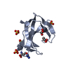
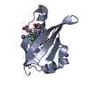
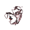
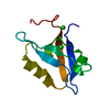
| Title | Crystal structure of the cell wall targeting domain of peptidylglycan hydrolase ALE-1 | ||||||
 Components Components | Cell Wall Targeting Domain of Glycylglycine Endopeptidase ALE-1 | ||||||
 Keywords Keywords | HYDROLASE / SH3b Domain / Cell Wall Targeting Domain / Lysostaphin / Peptidoglycan Hydrolase / Glycylglycine Endopeptidase | ||||||
| Function / homology |  Function and homology information Function and homology informationlysostaphin / cell wall organization / metalloendopeptidase activity / proteolysis / extracellular region / metal ion binding Similarity search - Function | ||||||
| Biological species |  Staphylococcus capitis (bacteria) Staphylococcus capitis (bacteria) | ||||||
| Method |  X-RAY DIFFRACTION / SYNCHROTRON / MOLECULAR REPLACEMENT / Resolution: 1.75 Å X-RAY DIFFRACTION / SYNCHROTRON / MOLECULAR REPLACEMENT / Resolution: 1.75 Å | ||||||
 Authors Authors | Lu, J.Z. / Fujiwara, T. / Komatsuzawa, H. / Sugai, M. / Sakon, J. | ||||||
 Citation Citation | Journal: J.Biol.Chem. / Year: 2006 Title: Cell Wall-targeting Domain of Glycylglycine Endopeptidase Distinguishes among Peptidoglycan Cross-bridges. Authors: Lu, J.Z. / Fujiwara, T. / Komatsuzawa, H. / Sugai, M. / Sakon, J. #1: Journal: J.Bacteriol. / Year: 1997Title: Purification and molecular characterization of glycylglycine endopeptidase produced by Staphylococcus capitis EPK1. Authors: Sugai, M. / Fujiwara, T. / Akiyama, T. / Ohara, M. / Komatsuzawa, H. / Inoue, S. / Suginaka, H. | ||||||
| History |
| ||||||
| Remark 999 | SEQUENCE FLAG-tag was added to the N-terminus |
- Structure visualization
| Structure viewer | Molecule: MolmilJmol/JSmol |
|---|
- Downloads & links
-Download
| PDBx/mmCIF format | 1r77.cif.gz | 56.5 KB | Display | PDBx/mmCIF format |
|---|---|---|---|---|
| PDB format | pdb1r77.ent.gz | 41.3 KB | Display | PDB format |
| PDBx/mmJSON format | 1r77.json.gz | Tree view | PDBx/mmJSON format | |
| Others |  Other downloads Other downloads |
-Validation report
| Arichive directory | https://data.pdbj.org/pub/pdb/validation_reports/r7/1r77ftp://data.pdbj.org/pub/pdb/validation_reports/r7/1r77 | HTTPS FTP |
|---|
-Related structure data
| Similar structure data |
|---|


-Links
 PDBj
PDBj- Assembly
| Deposited unit | 
| ||||||||
|---|---|---|---|---|---|---|---|---|---|
| 1 | 
| ||||||||
| 2 | 
| ||||||||
| Unit cell |
|
-Components
| #1: Protein | Mass: 11759.253 Da / Num. of mol.: 2 / Fragment: Cell Wall Targeting Domain / Mutation: FLAG-tag added at N-terminus Source method: isolated from a genetically manipulated source Source: (gene. exp.) Staphylococcus capitis (bacteria) / Strain: EPK1 / Plasmid: pTF479 / Production host: #2: Water | ChemComp-HOH / |  Mass: 18.015 Da / Num. of mol.: 221 / Source method: isolated from a natural source / Formula: H2O Mass: 18.015 Da / Num. of mol.: 221 / Source method: isolated from a natural source / Formula: H2O |
|---|
-Experimental details
-Experiment
| Experiment | Method: X-RAY DIFFRACTION / Number of used crystals: 4 |
|---|
- Sample preparation
| Crystal | Density Matthews: 2.39 Å3/Da / Density % sol: 48.58 % |
|---|---|
| Crystal grow | Temperature: 297 K / Method: vapor diffusion, hanging drop / pH: 6.5 Details: PEG 3350, sodium acetate, MES pH 6.5 or HEPES pH 7.5, VAPOR DIFFUSION, HANGING DROP, temperature 297K |
-Data collection
| Diffraction |
| ||||||||||||||||||
|---|---|---|---|---|---|---|---|---|---|---|---|---|---|---|---|---|---|---|---|
| Diffraction source |
| ||||||||||||||||||
| Detector |
| ||||||||||||||||||
| Radiation |
| ||||||||||||||||||
| Radiation wavelength |
| ||||||||||||||||||
| Reflection | Resolution: 1.75→20 Å / Num. all: 23430 / Num. obs: 22012 / % possible obs: 93.9 % / Observed criterion σ(F): 2 / Observed criterion σ(I): 2 / Biso Wilson estimate: 13.5 Å2 | ||||||||||||||||||
| Reflection shell | Resolution: 1.75→1.81 Å / % possible all: 98.9 |

- Processing
| Software |
| ||||||||||||||||||||||||||||||||||||
|---|---|---|---|---|---|---|---|---|---|---|---|---|---|---|---|---|---|---|---|---|---|---|---|---|---|---|---|---|---|---|---|---|---|---|---|---|---|
| Refinement | Method to determine structure: MOLECULAR REPLACEMENT Starting model: N-terminus truncated SeMet MAD structure Resolution: 1.75→17.5 Å / Rfactor Rfree error: 0.007 / Data cutoff high absF: 250222.27 / Data cutoff low absF: 0 / Isotropic thermal model: RESTRAINED / Cross valid method: THROUGHOUT / σ(F): 0
| ||||||||||||||||||||||||||||||||||||
| Solvent computation | Solvent model: FLAT MODEL / Bsol: 53.2203 Å2 / ksol: 0.389424 e/Å3 | ||||||||||||||||||||||||||||||||||||
| Displacement parameters | Biso mean: 19.5 Å2
| ||||||||||||||||||||||||||||||||||||
| Refine analyze |
| ||||||||||||||||||||||||||||||||||||
| Refinement step | Cycle: LAST / Resolution: 1.75→17.5 Å
| ||||||||||||||||||||||||||||||||||||
| Refine LS restraints |
| ||||||||||||||||||||||||||||||||||||
| LS refinement shell | Resolution: 1.75→1.86 Å / Rfactor Rfree error: 0.023 / Total num. of bins used: 6
| ||||||||||||||||||||||||||||||||||||
| Xplor file |
|
