 Movie
Movie Controller

[English] 日本語
 Yorodumi
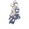
- PDB-1r3f: Crystal Structure of tRNA Pseudouridine Synthase TruB and Its RNA... -
+ Open data

- Basic information
| Entry | Database: PDB / ID: 1r3f | ||||||
|---|---|---|---|---|---|---|---|
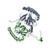
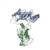
| Title | Crystal Structure of tRNA Pseudouridine Synthase TruB and Its RNA Complex: RNA-protein Recognition Through a Combination of Rigid Docking and Induced Fit | ||||||
 Components Components | tRNA pseudouridine synthase B | ||||||
 Keywords Keywords | LYASE / RNA modification / pseudouridylation | ||||||
| Function / homology |  Function and homology information Function and homology informationtRNA folding / tRNA pseudouridine synthase activity / tRNA pseudouridine55 synthase / tRNA pseudouridine(55) synthase activity / tRNA pseudouridine synthesis / mRNA pseudouridine synthesis / pseudouridine synthase activity / tRNA modification / tRNA binding / cytosol Similarity search - Function | ||||||
| Biological species |  | ||||||
| Method |  X-RAY DIFFRACTION / SYNCHROTRON / MAD / Resolution: 1.85 Å X-RAY DIFFRACTION / SYNCHROTRON / MAD / Resolution: 1.85 Å | ||||||
 Authors Authors | Pan, H. / Agarwalla, S. / Moustakas, D.T. / Finer-Moore, J. / Stroud, R.M. | ||||||
 Citation Citation | Journal: Proc.Natl.Acad.Sci.USA / Year: 2003 Title: Structure of tRNA Pseudouridine Synthase TruB and Its RNA Complex: RNA Recognition Through a Combination of Rigid Docking and Induced Fit Authors: Pan, H. / Agarwalla, S. / Moustakas, D.T. / Finer-Moore, J. / Stroud, R.M. | ||||||
| History |
|
- Structure visualization
| Structure viewer | Molecule: MolmilJmol/JSmol |
|---|
- Downloads & links
-Download
| PDBx/mmCIF format | 1r3f.cif.gz | 62.6 KB | Display | PDBx/mmCIF format |
|---|---|---|---|---|
| PDB format | pdb1r3f.ent.gz | 47.2 KB | Display | PDB format |
| PDBx/mmJSON format | 1r3f.json.gz | Tree view | PDBx/mmJSON format | |
| Others |  Other downloads Other downloads |
-Validation report
| Arichive directory | https://data.pdbj.org/pub/pdb/validation_reports/r3/1r3fftp://data.pdbj.org/pub/pdb/validation_reports/r3/1r3f | HTTPS FTP |
|---|
-Related structure data


-Links
 PDBj
PDBj
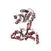
- Assembly
| Deposited unit | 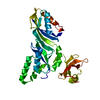
| ||||||||
|---|---|---|---|---|---|---|---|---|---|
| 1 |
| ||||||||
| Unit cell |
|
-Components
| #1: Protein | Mass: 35132.043 Da / Num. of mol.: 1 Source method: isolated from a genetically manipulated source Source: (gene. exp.) |
|---|
-Experimental details
-Experiment
| Experiment | Method: X-RAY DIFFRACTION / Number of used crystals: 1 |
|---|
- Sample preparation
| Crystal | Density Matthews: 2.09 Å3/Da / Density % sol: 41.07 % | ||||||||||||||||||||||||||||||||||||||||||||||||
|---|---|---|---|---|---|---|---|---|---|---|---|---|---|---|---|---|---|---|---|---|---|---|---|---|---|---|---|---|---|---|---|---|---|---|---|---|---|---|---|---|---|---|---|---|---|---|---|---|---|
| Crystal grow | Method: vapor diffusion, hanging drop / pH: 6.5 Details: PEG 4K, magnesium sulfate, sodium cacodylate, pH 6.5, VAPOR DIFFUSION, HANGING DROP | ||||||||||||||||||||||||||||||||||||||||||||||||
| Crystal grow | *PLUS pH: 7.5 / Method: vapor diffusion | ||||||||||||||||||||||||||||||||||||||||||||||||
| Components of the solutions | *PLUS
|
-Data collection
| Diffraction | Mean temperature: 298 K | ||||||||||||
|---|---|---|---|---|---|---|---|---|---|---|---|---|---|
| Diffraction source | Source: SYNCHROTRON / Site: ALS  / Beamline: 5.0.2 / Wavelength: 0.97926, 0.97942, 0.9610 / Beamline: 5.0.2 / Wavelength: 0.97926, 0.97942, 0.9610 | ||||||||||||
| Detector | Type: ADSC QUANTUM 4 / Detector: CCD / Date: Jun 10, 2001 / Details: mirrors | ||||||||||||
| Radiation | Monochromator: Double-crystal, Si(111) / Protocol: MAD / Monochromatic (M) / Laue (L): M / Scattering type: x-ray | ||||||||||||
| Radiation wavelength |
| ||||||||||||
| Reflection | Resolution: 1.85→40 Å / Num. all: 24806 / Num. obs: 24806 / % possible obs: 100 % / Observed criterion σ(F): 2 / Observed criterion σ(I): 1 / Redundancy: 9.97 % / Rmerge(I) obs: 0.069 / Rsym value: 0.069 / Net I/σ(I): 23.7 | ||||||||||||
| Reflection shell | Resolution: 1.85→1.96 Å / Rmerge(I) obs: 0.47 / Mean I/σ(I) obs: 4.9 / Rsym value: 0.47 / % possible all: 100 | ||||||||||||
| Reflection | *PLUS Num. measured all: 426647 | ||||||||||||
| Reflection shell | *PLUS % possible obs: 100 % / Rmerge(I) obs: 0.47 |
- Processing
| Software |
| ||||||||||||||||
|---|---|---|---|---|---|---|---|---|---|---|---|---|---|---|---|---|---|
| Refinement | Method to determine structure: MAD / Resolution: 1.85→50 Å / σ(F): 1.85 / Stereochemistry target values: Engh & Huber
| ||||||||||||||||
| Refinement step | Cycle: LAST / Resolution: 1.85→50 Å
| ||||||||||||||||
| Refine LS restraints |
| ||||||||||||||||
| LS refinement shell | Resolution: 1.85→1.96 Å / Rfactor Rfree: 0.257 / Rfactor Rwork: 0.228 | ||||||||||||||||
| Refinement | *PLUS Lowest resolution: 40 Å / Num. reflection obs: 46517 | ||||||||||||||||
| Solvent computation | *PLUS | ||||||||||||||||
| Displacement parameters | *PLUS | ||||||||||||||||
| Refine LS restraints | *PLUS
|
