 Movie
Movie Controller


+ Open data

- Basic information
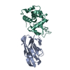
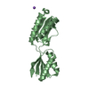
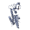
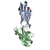
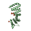
| Entry | Database: PDB / ID: 1r0d | ||||||
|---|---|---|---|---|---|---|---|
| Title | HIP1R THATCH DOMAIN CORE | ||||||
 Components Components | Huntingtin Interacting Protein 12 | ||||||
 Keywords Keywords | STRUCTURAL PROTEIN / Endocytosis / Actin-binding / Structural Genomics / PSI / Protein Structure Initiative / Midwest Center for Structural Genomics / MCSG | ||||||
| Function / homology |  Function and homology information Function and homology informationpositive regulation of clathrin coat assembly / digestive system development / regulation of clathrin-dependent endocytosis / regulation of gastric acid secretion / clathrin light chain binding / positive regulation of platelet-derived growth factor receptor-beta signaling pathway / postsynapse organization / negative regulation of actin filament polymerization / postsynaptic endocytic zone / negative regulation of Arp2/3 complex-mediated actin nucleation ...positive regulation of clathrin coat assembly / digestive system development / regulation of clathrin-dependent endocytosis / regulation of gastric acid secretion / clathrin light chain binding / positive regulation of platelet-derived growth factor receptor-beta signaling pathway / postsynapse organization / negative regulation of actin filament polymerization / postsynaptic endocytic zone / negative regulation of Arp2/3 complex-mediated actin nucleation / clathrin-coated vesicle membrane / clathrin coat assembly / clathrin-cargo adaptor activity / positive regulation of mitochondrial outer membrane permeabilization involved in apoptotic signaling pathway / phosphatidylinositol-3,4-bisphosphate binding / phosphatidylinositol-3,5-bisphosphate binding / clathrin-coated vesicle / cortical actin cytoskeleton / clathrin binding / Golgi Associated Vesicle Biogenesis / phosphatidylinositol-3,4,5-trisphosphate binding / regulation of endocytosis / positive regulation of epidermal growth factor receptor signaling pathway / phosphatidylinositol-4,5-bisphosphate binding / clathrin-coated pit / phosphatidylinositol binding / intrinsic apoptotic signaling pathway / receptor-mediated endocytosis / dendrite cytoplasm / actin filament organization / regulation of actin cytoskeleton organization / synaptic membrane / SH3 domain binding / ruffle membrane / endocytosis / actin filament binding / Clathrin-mediated endocytosis / regulation of apoptotic process / dendritic spine / apical plasma membrane / postsynaptic density / protein stabilization / protein heterodimerization activity / neuronal cell body / negative regulation of apoptotic process / perinuclear region of cytoplasm / glutamatergic synapse / protein homodimerization activity / mitochondrion / identical protein binding / plasma membrane / cytosol Similarity search - Function | ||||||
| Biological species |  Homo sapiens (human) Homo sapiens (human) | ||||||
| Method |  X-RAY DIFFRACTION / SYNCHROTRON / MAD / Resolution: 1.9 Å X-RAY DIFFRACTION / SYNCHROTRON / MAD / Resolution: 1.9 Å | ||||||
 Authors Authors | Brett, T.J. / Fremont, D.H. / Midwest Center for Structural Genomics (MCSG) | ||||||
 Citation Citation | Journal: Nat.Struct.Mol.Biol. / Year: 2006 Title: Structural definition of the F-actin-binding THATCH domain from HIP1R Authors: Brett, T.J. / Legendre-Guillemin, V. / McPherson, P.S. / Fremont, D.H. | ||||||
| History |
|
- Structure visualization
| Structure viewer | Molecule: MolmilJmol/JSmol |
|---|
- Downloads & links
-Download
| PDBx/mmCIF format | 1r0d.cif.gz | 317 KB | Display | PDBx/mmCIF format |
|---|---|---|---|---|
| PDB format | pdb1r0d.ent.gz | 260 KB | Display | PDB format |
| PDBx/mmJSON format | 1r0d.json.gz | Tree view | PDBx/mmJSON format | |
| Others |  Other downloads Other downloads |
-Validation report
| Arichive directory | https://data.pdbj.org/pub/pdb/validation_reports/r0/1r0dftp://data.pdbj.org/pub/pdb/validation_reports/r0/1r0d | HTTPS FTP |
|---|
-Related structure data
| Similar structure data | |
|---|---|
| Other databases |


-Links
 PDBj
PDBj


- Assembly
| Deposited unit | 
| ||||||||||
|---|---|---|---|---|---|---|---|---|---|---|---|
| 1 | 
| ||||||||||
| 2 | 
| ||||||||||
| 3 | 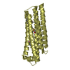
| ||||||||||
| 4 | 
| ||||||||||
| 5 | 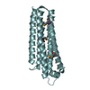
| ||||||||||
| 6 | 
| ||||||||||
| 7 | 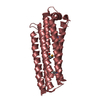
| ||||||||||
| 8 | 
| ||||||||||
| Unit cell |
|
-Components
| #1: Protein | Mass: 22609.607 Da / Num. of mol.: 8 / Fragment: C-TERMINAL THATCH DOMAIN, RESIDUES 771-971 Source method: isolated from a genetically manipulated source Source: (gene. exp.) Homo sapiens (human) / Gene: HIP1R, HIP12, KIAA0655 / Plasmid: PGEX-4T1 / Species (production host): Escherichia coli / Production host:  #2: Water | ChemComp-HOH / |  Mass: 18.015 Da / Num. of mol.: 1149 / Source method: isolated from a natural source / Formula: H2O Mass: 18.015 Da / Num. of mol.: 1149 / Source method: isolated from a natural source / Formula: H2OHas protein modification | Y | |
|---|
-Experimental details
-Experiment
| Experiment | Method: X-RAY DIFFRACTION / Number of used crystals: 1 |
|---|
- Sample preparation
| Crystal | Density Matthews: 2.76 Å3/Da / Density % sol: 58 % |
|---|---|
| Crystal grow | Temperature: 293 K / Method: vapor diffusion, hanging drop / pH: 4.8 Details: 1.75 M ammonium sulfate, 2.5% ethylene glycol, pH 4.8, VAPOR DIFFUSION, HANGING DROP, temperature 293K |
-Data collection
| Diffraction |
| ||||||||||||||||||
|---|---|---|---|---|---|---|---|---|---|---|---|---|---|---|---|---|---|---|---|
| Diffraction source |
| ||||||||||||||||||
| Detector |
| ||||||||||||||||||
| Radiation |
| ||||||||||||||||||
| Radiation wavelength |
| ||||||||||||||||||
| Reflection | Resolution: 1.9→50 Å / Num. all: 147170 / Num. obs: 147170 / % possible obs: 95.7 % / Observed criterion σ(F): 0 / Observed criterion σ(I): 0 / Biso Wilson estimate: 23.3 Å2 / Rmerge(I) obs: 0.043 / Rsym value: 0.043 / Net I/σ(I): 23.5 | ||||||||||||||||||
| Reflection shell | Resolution: 1.9→1.97 Å / Rmerge(I) obs: 0.433 / Mean I/σ(I) obs: 2.4 / Rsym value: 0.433 / % possible all: 95.2 |

- Processing
| Software |
| ||||||||||||||||||||||||||||||||||||
|---|---|---|---|---|---|---|---|---|---|---|---|---|---|---|---|---|---|---|---|---|---|---|---|---|---|---|---|---|---|---|---|---|---|---|---|---|---|
| Refinement | Method to determine structure: MAD / Resolution: 1.9→19.88 Å / Rfactor Rfree error: 0.002 / Isotropic thermal model: RESTRAINED / Cross valid method: THROUGHOUT / σ(F): 0 / Stereochemistry target values: Engh & Huber
| ||||||||||||||||||||||||||||||||||||
| Solvent computation | Solvent model: FLAT MODEL / Bsol: 63.952 Å2 / ksol: 0.362315 e/Å3 | ||||||||||||||||||||||||||||||||||||
| Displacement parameters | Biso mean: 36.9 Å2
| ||||||||||||||||||||||||||||||||||||
| Refine analyze |
| ||||||||||||||||||||||||||||||||||||
| Refinement step | Cycle: LAST / Resolution: 1.9→19.88 Å
| ||||||||||||||||||||||||||||||||||||
| Refine LS restraints |
| ||||||||||||||||||||||||||||||||||||
| LS refinement shell | Resolution: 1.9→2.02 Å / Rfactor Rfree error: 0.009 / Total num. of bins used: 6
| ||||||||||||||||||||||||||||||||||||
| Xplor file |
|
