 Movie
Movie Controller


+ Open data

- Basic information
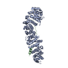
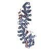
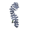
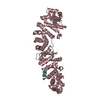
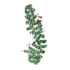
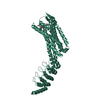
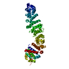
| Entry | Database: PDB / ID: 1qz7 | ||||||
|---|---|---|---|---|---|---|---|
| Title | Beta-catenin binding domain of Axin in complex with beta-catenin | ||||||
 Components Components |
| ||||||
 Keywords Keywords | CELL ADHESION / Beta-catenin / Axin / protein-protein complex | ||||||
| Function / homology |  Function and homology information Function and homology informationcanonical Wnt signaling pathway involved in mesenchymal stem cell differentiation / positive regulation of heparan sulfate proteoglycan biosynthetic process / lung induction / positive regulation of branching involved in lung morphogenesis / cranial ganglion development / renal vesicle formation / renal inner medulla development / renal outer medulla development / nephron tubule formation / beta-catenin-ICAT complex ...canonical Wnt signaling pathway involved in mesenchymal stem cell differentiation / positive regulation of heparan sulfate proteoglycan biosynthetic process / lung induction / positive regulation of branching involved in lung morphogenesis / cranial ganglion development / renal vesicle formation / renal inner medulla development / renal outer medulla development / nephron tubule formation / beta-catenin-ICAT complex / CDH11 homotypic and heterotypic interactions / genitalia morphogenesis / neural plate development / metanephros morphogenesis / glial cell fate determination / Regulation of CDH19 Expression and Function / astrocyte-dopaminergic neuron signaling / oviduct development / beta-catenin-TCF7L2 complex / regulation of nephron tubule epithelial cell differentiation / regulation of timing of anagen / negative regulation of mitotic cell cycle, embryonic / regulation of secondary heart field cardioblast proliferation / Binding of TCF/LEF:CTNNB1 to target gene promoters / central nervous system vasculogenesis / negative regulation of mesenchymal to epithelial transition involved in metanephros morphogenesis / embryonic skeletal limb joint morphogenesis / regulation of centriole-centriole cohesion / RUNX3 regulates WNT signaling / regulation of centromeric sister chromatid cohesion / Regulation of CDH11 function / acinar cell differentiation / embryonic axis specification / lens morphogenesis in camera-type eye / Scrib-APC-beta-catenin complex / regulation of fibroblast proliferation / beta-catenin-TCF complex / Specification of the neural plate border / neuron fate determination / cell development / synaptic vesicle clustering / endodermal cell fate commitment / Formation of the nephric duct / proximal/distal pattern formation / dorsal root ganglion development / positive regulation of fibroblast growth factor receptor signaling pathway / lung epithelial cell differentiation / endothelial tube morphogenesis / dorsal/ventral axis specification / sympathetic ganglion development / positive regulation of endothelial cell differentiation / layer formation in cerebral cortex / mesenchymal to epithelial transition / presynaptic active zone cytoplasmic component / positive regulation of skeletal muscle tissue development / fungiform papilla formation / positive regulation of determination of dorsal identity / ectoderm development / regulation of protein localization to cell surface / fascia adherens / hindbrain development / positive regulation of myoblast proliferation / embryonic foregut morphogenesis / detection of muscle stretch / positive regulation of odontoblast differentiation / smooth muscle cell differentiation / mesenchymal cell proliferation involved in lung development / hair cell differentiation / alpha-catenin binding / cellular response to indole-3-methanol / histone methyltransferase binding / regulation of epithelial to mesenchymal transition / regulation of calcium ion import / Germ layer formation at gastrulation / positive regulation of homotypic cell-cell adhesion / negative regulation of oligodendrocyte differentiation / establishment of blood-retinal barrier / apicolateral plasma membrane / epithelial cell differentiation involved in prostate gland development / epithelial cell proliferation involved in prostate gland development / positive regulation of epithelial cell proliferation involved in prostate gland development / male genitalia development / flotillin complex / cranial skeletal system development / cell-cell adhesion mediated by cadherin / Formation of definitive endoderm / regulation of smooth muscle cell proliferation / lung-associated mesenchyme development / establishment of blood-brain barrier / Formation of axial mesoderm / beta-catenin destruction complex / negative regulation of protein sumoylation / midbrain dopaminergic neuron differentiation / Apoptotic cleavage of cell adhesion proteins / catenin complex / LRR FLII-interacting protein 1 (LRRFIP1) activates type I IFN production / embryonic brain development / positive regulation of blood vessel branching / embryonic heart tube development / pancreas development Similarity search - Function | ||||||
| Biological species |  Homo sapiens (human) Homo sapiens (human) | ||||||
| Method |  X-RAY DIFFRACTION / SYNCHROTRON / MOLECULAR REPLACEMENT / Resolution: 2.2 Å X-RAY DIFFRACTION / SYNCHROTRON / MOLECULAR REPLACEMENT / Resolution: 2.2 Å | ||||||
 Authors Authors | Xing, Y. / Clements, W.K. / Kimelman, D. / Xu, W. | ||||||
 Citation Citation | Journal: GENES DEV. / Year: 2003 Title: Crystal structure of a beta-catenin/Axin complex suggests a mechanism for the {beta}-catenin destruction complex Authors: Xing, Y. / Clements, W.K. / Kimelman, D. / Xu, W. | ||||||
| History |
|
- Structure visualization
| Structure viewer | Molecule: MolmilJmol/JSmol |
|---|
- Downloads & links
-Download
| PDBx/mmCIF format | 1qz7.cif.gz | 115.3 KB | Display | PDBx/mmCIF format |
|---|---|---|---|---|
| PDB format | pdb1qz7.ent.gz | 87.4 KB | Display | PDB format |
| PDBx/mmJSON format | 1qz7.json.gz | Tree view | PDBx/mmJSON format | |
| Others |  Other downloads Other downloads |
-Validation report
| Arichive directory | https://data.pdbj.org/pub/pdb/validation_reports/qz/1qz7ftp://data.pdbj.org/pub/pdb/validation_reports/qz/1qz7 | HTTPS FTP |
|---|
-Related structure data
| Related structure data |  1i7xS S: Starting model for refinement |
|---|---|
| Similar structure data |


-Links
 PDBj
PDBj


- Assembly
| Deposited unit | 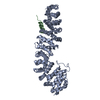
| ||||||||
|---|---|---|---|---|---|---|---|---|---|
| 1 |
| ||||||||
| Unit cell |
|
-Components
| #1: Protein | Mass: 58211.438 Da / Num. of mol.: 1 / Fragment: Armadillo repeat region Source method: isolated from a genetically manipulated source Source: (gene. exp.) Homo sapiens (human) / Gene: CTNNB1 / Plasmid: PGEX / Production host:  |
|---|---|
| #2: Protein | Mass: 7756.588 Da / Num. of mol.: 1 / Fragment: Beta-catenin binding domain Source method: isolated from a genetically manipulated source Source: (gene. exp.) |
| #3: Water | ChemComp-HOH /  Mass: 18.015 Da / Num. of mol.: 31 / Source method: isolated from a natural source / Formula: H2O Mass: 18.015 Da / Num. of mol.: 31 / Source method: isolated from a natural source / Formula: H2O |
-Experimental details
-Experiment
| Experiment | Method: X-RAY DIFFRACTION / Number of used crystals: 1 |
|---|
- Sample preparation
| Crystal | Density Matthews: 2.43 Å3/Da / Density % sol: 49.45 % | |||||||||||||||||||||||||||||||||||||||||||||||||||||||||||||||
|---|---|---|---|---|---|---|---|---|---|---|---|---|---|---|---|---|---|---|---|---|---|---|---|---|---|---|---|---|---|---|---|---|---|---|---|---|---|---|---|---|---|---|---|---|---|---|---|---|---|---|---|---|---|---|---|---|---|---|---|---|---|---|---|---|
| Crystal grow | Temperature: 298 K / Method: vapor diffusion, hanging drop / pH: 5.6 Details: PEG 6000, sodium citrate, isopropanol, pH 5.6, VAPOR DIFFUSION, HANGING DROP, temperature 298K | |||||||||||||||||||||||||||||||||||||||||||||||||||||||||||||||
| Crystal grow | *PLUS Temperature: 20 ℃ / pH: 8.5 / Method: vapor diffusion, hanging drop | |||||||||||||||||||||||||||||||||||||||||||||||||||||||||||||||
| Components of the solutions | *PLUS
|
-Data collection
| Diffraction | Mean temperature: 110 K |
|---|---|
| Diffraction source | Source: SYNCHROTRON / Site: ALS  / Beamline: 5.0.1 / Wavelength: 1 Å / Beamline: 5.0.1 / Wavelength: 1 Å |
| Detector | Type: ADSC QUANTUM 4 / Detector: CCD / Date: Dec 5, 2002 |
| Radiation | Monochromator: Si(220) / Protocol: SINGLE WAVELENGTH / Monochromatic (M) / Laue (L): M / Scattering type: x-ray |
| Radiation wavelength | Wavelength: 1 Å / Relative weight: 1 |
| Reflection | Resolution: 2.1→50 Å / Num. all: 37253 / Num. obs: 37253 / % possible obs: 99.8 % / Redundancy: 3.7 % / Biso Wilson estimate: 40.7 Å2 / Rmerge(I) obs: 0.072 / Rsym value: 0.072 / Net I/σ(I): 17.8 |
| Reflection shell | Resolution: 2.1→2.18 Å / Redundancy: 3.1 % / Rmerge(I) obs: 0.702 / Mean I/σ(I) obs: 1.8 / Num. unique all: 3651 / Rsym value: 0.702 / % possible all: 99.3 |
| Reflection | *PLUS Highest resolution: 2.2 Å / Lowest resolution: 50 Å |
| Reflection shell | *PLUS % possible obs: 99.9 % / Rmerge(I) obs: 0.529 / Mean I/σ(I) obs: 2.7 |
- Processing
| Software |
| ||||||||||||||||||||||||||||||||||||
|---|---|---|---|---|---|---|---|---|---|---|---|---|---|---|---|---|---|---|---|---|---|---|---|---|---|---|---|---|---|---|---|---|---|---|---|---|---|
| Refinement | Method to determine structure: MOLECULAR REPLACEMENT Starting model: PDB entry 1I7X Resolution: 2.2→42.21 Å / Rfactor Rfree error: 0.007 / Data cutoff high absF: 267694.88 / Data cutoff high rms absF: 267694.88 / Data cutoff low absF: 0 / Isotropic thermal model: RESTRAINED / Cross valid method: THROUGHOUT / σ(F): 1 / Stereochemistry target values: Engh & Huber
| ||||||||||||||||||||||||||||||||||||
| Solvent computation | Solvent model: FLAT MODEL / Bsol: 45.402 Å2 / ksol: 0.345268 e/Å3 | ||||||||||||||||||||||||||||||||||||
| Displacement parameters | Biso mean: 54.1 Å2
| ||||||||||||||||||||||||||||||||||||
| Refine analyze |
| ||||||||||||||||||||||||||||||||||||
| Refinement step | Cycle: LAST / Resolution: 2.2→42.21 Å
| ||||||||||||||||||||||||||||||||||||
| Refine LS restraints |
| ||||||||||||||||||||||||||||||||||||
| LS refinement shell | Resolution: 2.2→2.34 Å / Rfactor Rfree error: 0.024 / Total num. of bins used: 6
| ||||||||||||||||||||||||||||||||||||
| Xplor file |
| ||||||||||||||||||||||||||||||||||||
| Refinement | *PLUS Highest resolution: 2.2 Å | ||||||||||||||||||||||||||||||||||||
| Solvent computation | *PLUS | ||||||||||||||||||||||||||||||||||||
| Displacement parameters | *PLUS | ||||||||||||||||||||||||||||||||||||
| Refine LS restraints | *PLUS
|
