 Movie
Movie Controller


+ Open data

- Basic information
| Entry | Database: PDB / ID: 1qyn | ||||||
|---|---|---|---|---|---|---|---|
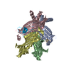
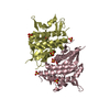
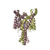
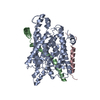
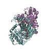
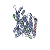
| Title | Crystal Structure of SecB from Escherichia coli | ||||||
 Components Components | Protein-export protein secB | ||||||
 Keywords Keywords | CHAPERONE / tetramer / Greek key beta sheet | ||||||
| Function / homology |  Function and homology information Function and homology information: / preprotein binding / : / protein transport by the Sec complex / protein targeting / protein tetramerization / : / intracellular protein localization / protein transport / protein folding / cytosol Similarity search - Function | ||||||
| Biological species |  | ||||||
| Method |  X-RAY DIFFRACTION / SYNCHROTRON / MOLECULAR REPLACEMENT / Resolution: 2.35 Å X-RAY DIFFRACTION / SYNCHROTRON / MOLECULAR REPLACEMENT / Resolution: 2.35 Å | ||||||
 Authors Authors | Dekker, C. / de Kruijff, B. / Gros, P. | ||||||
 Citation Citation | Journal: J.Struct.Biol. / Year: 2003 Title: Crystal structure of SecB from Escherichia coli Authors: Dekker, C. / de Kruijff, B. / Gros, P. | ||||||
| History |
|
- Structure visualization
| Structure viewer | Molecule: MolmilJmol/JSmol |
|---|
- Downloads & links
-Download
| PDBx/mmCIF format | 1qyn.cif.gz | 114 KB | Display | PDBx/mmCIF format |
|---|---|---|---|---|
| PDB format | pdb1qyn.ent.gz | 90.5 KB | Display | PDB format |
| PDBx/mmJSON format | 1qyn.json.gz | Tree view | PDBx/mmJSON format | |
| Others |  Other downloads Other downloads |
-Validation report
| Arichive directory | https://data.pdbj.org/pub/pdb/validation_reports/qy/1qynftp://data.pdbj.org/pub/pdb/validation_reports/qy/1qyn | HTTPS FTP |
|---|
-Related structure data
| Related structure data | 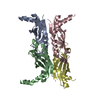 1fx3S S: Starting model for refinement |
|---|---|
| Similar structure data |


-Links
 PDBj
PDBj
- Assembly
| Deposited unit | 
| ||||||||
|---|---|---|---|---|---|---|---|---|---|
| 1 |
| ||||||||
| Unit cell |
| ||||||||
| Details | The asymmetric unit contains one tetramer which is the biological assembly of this chaperone |
-Components
| #1: Protein | Mass: 17101.098 Da / Num. of mol.: 4 Source method: isolated from a genetically manipulated source Source: (gene. exp.) #2: Water | ChemComp-HOH / |  Mass: 18.015 Da / Num. of mol.: 113 / Source method: isolated from a natural source / Formula: H2O Mass: 18.015 Da / Num. of mol.: 113 / Source method: isolated from a natural source / Formula: H2O |
|---|
-Experimental details
-Experiment
| Experiment | Method: X-RAY DIFFRACTION / Number of used crystals: 1 |
|---|
- Sample preparation
| Crystal | Density Matthews: 2.64 Å3/Da / Density % sol: 53.43 % | ||||||||||||||||||||||||
|---|---|---|---|---|---|---|---|---|---|---|---|---|---|---|---|---|---|---|---|---|---|---|---|---|---|
| Crystal grow | Temperature: 277 K / Method: vapor diffusion, hanging drop / pH: 7 Details: calcium chloride, MPD, pH 7, VAPOR DIFFUSION, HANGING DROP, temperature 277K | ||||||||||||||||||||||||
| Crystal grow | *PLUS Temperature: 4 ℃ / Method: vapor diffusion, hanging drop / Details: Dekker, C., (2003) J.Struct.Biol., 144, 313. | ||||||||||||||||||||||||
| Components of the solutions | *PLUS
|
-Data collection
| Diffraction | Mean temperature: 100 K |
|---|---|
| Diffraction source | Source: SYNCHROTRON / Site: EMBL/DESY, HAMBURG  / Beamline: X11 / Wavelength: 0.98 Å / Beamline: X11 / Wavelength: 0.98 Å |
| Detector | Type: MARRESEARCH / Detector: IMAGE PLATE / Date: Mar 8, 1997 |
| Radiation | Monochromator: graphite / Protocol: SINGLE WAVELENGTH / Monochromatic (M) / Laue (L): M / Scattering type: x-ray |
| Radiation wavelength | Wavelength: 0.98 Å / Relative weight: 1 |
| Reflection | Resolution: 2.35→29.61 Å / Num. all: 31445 / Num. obs: 30156 / % possible obs: 95.9 % / Observed criterion σ(F): 2 / Observed criterion σ(I): 2 / Redundancy: 13.9 % / Biso Wilson estimate: 46.7 Å2 / Rmerge(I) obs: 0.047 / Net I/σ(I): 28.6 |
| Reflection shell | Resolution: 2.35→2.5 Å / Rmerge(I) obs: 0.219 / Mean I/σ(I) obs: 5 / % possible all: 96.7 |
| Reflection | *PLUS Num. obs: 33605 / % possible obs: 95.8 % / Num. measured all: 465596 / Rmerge(I) obs: 0.045 |
- Processing
| Software |
| ||||||||||||||||||||||||||||||||||||
|---|---|---|---|---|---|---|---|---|---|---|---|---|---|---|---|---|---|---|---|---|---|---|---|---|---|---|---|---|---|---|---|---|---|---|---|---|---|
| Refinement | Method to determine structure: MOLECULAR REPLACEMENT Starting model: PDB entry 1FX3 Resolution: 2.35→29.61 Å / Rfactor Rfree error: 0.005 / Data cutoff high absF: 131500.99 / Data cutoff high rms absF: 131500.99 / Data cutoff low absF: 0 / Isotropic thermal model: RESTRAINED / Cross valid method: THROUGHOUT / σ(F): 0 / Stereochemistry target values: Engh & Huber
| ||||||||||||||||||||||||||||||||||||
| Solvent computation | Solvent model: FLAT MODEL / Bsol: 41.8554 Å2 / ksol: 0.334665 e/Å3 | ||||||||||||||||||||||||||||||||||||
| Displacement parameters | Biso mean: 54 Å2
| ||||||||||||||||||||||||||||||||||||
| Refine analyze |
| ||||||||||||||||||||||||||||||||||||
| Refinement step | Cycle: LAST / Resolution: 2.35→29.61 Å
| ||||||||||||||||||||||||||||||||||||
| Refine LS restraints |
| ||||||||||||||||||||||||||||||||||||
| LS refinement shell | Resolution: 2.35→2.5 Å / Rfactor Rfree error: 0.016 / Total num. of bins used: 6
| ||||||||||||||||||||||||||||||||||||
| Xplor file |
| ||||||||||||||||||||||||||||||||||||
| Software | *PLUS Name: CNS / Classification: refinement | ||||||||||||||||||||||||||||||||||||
| Refinement | *PLUS Lowest resolution: 29.6 Å / % reflection Rfree: 10 % / Rfactor Rwork: 0.235 | ||||||||||||||||||||||||||||||||||||
| Solvent computation | *PLUS | ||||||||||||||||||||||||||||||||||||
| Displacement parameters | *PLUS | ||||||||||||||||||||||||||||||||||||
| Refine LS restraints | *PLUS
|
