 Movie
Movie Controller

[English] 日本語
 Yorodumi
- PDB-1qtw: HIGH-RESOLUTION CRYSTAL STRUCTURE OF THE ESCHERICHIA COLI DNA REP... -
+ Open data

- Basic information
| Entry | Database: PDB / ID: 1qtw | ||||||
|---|---|---|---|---|---|---|---|
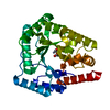
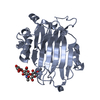
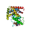
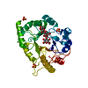
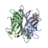
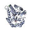
| Title | HIGH-RESOLUTION CRYSTAL STRUCTURE OF THE ESCHERICHIA COLI DNA REPAIR ENZYME ENDONUCLEASE IV | ||||||
 Components Components | ENDONUCLEASE IV | ||||||
 Keywords Keywords | HYDROLASE / DNA REPAIR ENZYME / TIM BARREL / TRINUCLEAR ZN CLUSTER | ||||||
| Function / homology |  Function and homology information Function and homology informationdeoxyribonuclease IV / deoxyribonuclease IV (phage-T4-induced) activity / phosphoric diester hydrolase activity / 3'-5'-DNA exonuclease activity / phosphatase activity / DNA-(apurinic or apyrimidinic site) endonuclease activity / base-excision repair / endonuclease activity / DNA repair / DNA binding ...deoxyribonuclease IV / deoxyribonuclease IV (phage-T4-induced) activity / phosphoric diester hydrolase activity / 3'-5'-DNA exonuclease activity / phosphatase activity / DNA-(apurinic or apyrimidinic site) endonuclease activity / base-excision repair / endonuclease activity / DNA repair / DNA binding / zinc ion binding / cytosol Similarity search - Function | ||||||
| Biological species |  | ||||||
| Method |  X-RAY DIFFRACTION / SYNCHROTRON / Resolution: 1.02 Å X-RAY DIFFRACTION / SYNCHROTRON / Resolution: 1.02 Å | ||||||
 Authors Authors | Hosfield, D.J. / Guan, Y. / Haas, B.J. / Cunningham, R.P. / Tainer, J.A. | ||||||
 Citation Citation | Journal: Cell(Cambridge,Mass.) / Year: 1999 Title: Structure of the DNA repair enzyme endonuclease IV and its DNA complex: double-nucleotide flipping at abasic sites and three-metal-ion catalysis. Authors: Hosfield, D.J. / Guan, Y. / Haas, B.J. / Cunningham, R.P. / Tainer, J.A. | ||||||
| History |
|
- Structure visualization
| Structure viewer | Molecule: MolmilJmol/JSmol |
|---|
- Downloads & links
-Download
| PDBx/mmCIF format | 1qtw.cif.gz | 184.3 KB | Display | PDBx/mmCIF format |
|---|---|---|---|---|
| PDB format | pdb1qtw.ent.gz | 148.2 KB | Display | PDB format |
| PDBx/mmJSON format | 1qtw.json.gz | Tree view | PDBx/mmJSON format | |
| Others |  Other downloads Other downloads |
-Validation report
| Summary document | 1qtw_validation.pdf.gz | 364.6 KB | Display | wwPDB validaton report |
|---|---|---|---|---|
| Full document | 1qtw_full_validation.pdf.gz | 369.6 KB | Display | |
| Data in XML | 1qtw_validation.xml.gz | 7.5 KB | Display | |
| Data in CIF | 1qtw_validation.cif.gz | 13 KB | Display | |
| Arichive directory | https://data.pdbj.org/pub/pdb/validation_reports/qt/1qtwftp://data.pdbj.org/pub/pdb/validation_reports/qt/1qtw | HTTPS FTP |
-Related structure data

-Links
 PDBj
PDBj
- Assembly
| Deposited unit | 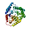
| ||||||||||
|---|---|---|---|---|---|---|---|---|---|---|---|
| 1 |
| ||||||||||
| Unit cell |
|
-Components
| #1: Protein | Mass: 31518.496 Da / Num. of mol.: 1 Source method: isolated from a genetically manipulated source Source: (gene. exp.) | ||
|---|---|---|---|
| #2: Chemical |   Mass: 65.409 Da / Num. of mol.: 3 / Source method: obtained synthetically / Formula: Zn Mass: 65.409 Da / Num. of mol.: 3 / Source method: obtained synthetically / Formula: Zn#3: Water | ChemComp-HOH / |  Mass: 18.015 Da / Num. of mol.: 367 / Source method: isolated from a natural source / Formula: H2O Mass: 18.015 Da / Num. of mol.: 367 / Source method: isolated from a natural source / Formula: H2O |
-Experimental details
-Experiment
| Experiment | Method: X-RAY DIFFRACTION / Number of used crystals: 1 |
|---|
- Sample preparation
| Crystal | Density Matthews: 2.23 Å3/Da / Density % sol: 44.89 % | |||||||||||||||
|---|---|---|---|---|---|---|---|---|---|---|---|---|---|---|---|---|
| Crystal grow | Temperature: 298 K / Method: vapor diffusion, sitting drop / pH: 6 Details: PEG 400, HEPES, ZNCL2, pH 6.0, VAPOR DIFFUSION, SITTING DROP at 298 K | |||||||||||||||
| Crystal grow | *PLUS pH: 6.5 / Method: vapor diffusion, hanging drop | |||||||||||||||
| Components of the solutions | *PLUS
|
-Data collection
| Diffraction | Mean temperature: 100 K |
|---|---|
| Diffraction source | Source: SYNCHROTRON / Site: SSRL  / Beamline: BL9-1 / Wavelength: 0.98 / Beamline: BL9-1 / Wavelength: 0.98 |
| Detector | Type: MARRESEARCH / Detector: IMAGE PLATE / Date: Mar 19, 1998 |
| Radiation | Protocol: SINGLE WAVELENGTH / Monochromatic (M) / Laue (L): M / Scattering type: x-ray |
| Radiation wavelength | Wavelength: 0.98 Å / Relative weight: 1 |
| Reflection | Resolution: 1.02→30 Å / Num. obs: 142898 / % possible obs: 96 % / Observed criterion σ(I): -3 / Redundancy: 3.26 % / Biso Wilson estimate: 12.6 Å2 / Rmerge(I) obs: 0.042 / Net I/σ(I): 13.2 |
| Reflection shell | Resolution: 1.02→1.04 Å / Redundancy: 2.2 % / Rmerge(I) obs: 0.436 / Mean I/σ(I) obs: 2.5 / % possible all: 83.2 |
| Reflection | *PLUS Highest resolution: 1.02 Å / Lowest resolution: 30 Å / Observed criterion σ(I): -3 / Redundancy: 3.26 % / Num. measured all: 465830 |
| Reflection shell | *PLUS Mean I/σ(I) obs: 2.5 |
- Processing
| Software |
| |||||||||||||||||||||||||||||||||
|---|---|---|---|---|---|---|---|---|---|---|---|---|---|---|---|---|---|---|---|---|---|---|---|---|---|---|---|---|---|---|---|---|---|---|
| Refinement | Resolution: 1.02→20 Å / Cross valid method: THROUGHOUT / σ(F): 0 / Stereochemistry target values: ENGH & HUBER
| |||||||||||||||||||||||||||||||||
| Refinement step | Cycle: LAST / Resolution: 1.02→20 Å
| |||||||||||||||||||||||||||||||||
| Refine LS restraints |
| |||||||||||||||||||||||||||||||||
| Software | *PLUS Name: SHELXL-97 / Classification: refinement | |||||||||||||||||||||||||||||||||
| Refinement | *PLUS Lowest resolution: 20 Å | |||||||||||||||||||||||||||||||||
| Solvent computation | *PLUS | |||||||||||||||||||||||||||||||||
| Displacement parameters | *PLUS |
