 Movie
Movie Controller

[English] 日本語
 Yorodumi
- PDB-1lqp: CRYSTAL STRUCTURE OF THE FOSFOMYCIN RESISTANCE PROTEIN (FOSA) CON... -
+ Open data

- Basic information
| Entry | Database: PDB / ID: 1lqp | ||||||
|---|---|---|---|---|---|---|---|
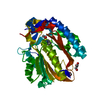
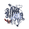
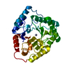
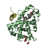
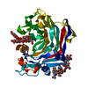
| Title | CRYSTAL STRUCTURE OF THE FOSFOMYCIN RESISTANCE PROTEIN (FOSA) CONTAINING BOUND SUBSTRATE | ||||||
 Components Components | PROBABLE FOSFOMYCIN RESISTANCE PROTEIN | ||||||
 Keywords Keywords | TRANSFERASE / potassium binding loop / manganese binding | ||||||
| Function / homology |  Function and homology information Function and homology informationglutathione transferase / glutathione transferase activity / response to antibiotic / metal ion binding / cytoplasm Similarity search - Function | ||||||
| Biological species |   Pseudomonas aeruginosa (bacteria) Pseudomonas aeruginosa (bacteria) | ||||||
| Method |  X-RAY DIFFRACTION / SYNCHROTRON / MAD / Resolution: 1.19 Å X-RAY DIFFRACTION / SYNCHROTRON / MAD / Resolution: 1.19 Å | ||||||
 Authors Authors | Rife, C.L. / Pharris, R.E. / Newcomer, M.E. / Armstrong, R.N. | ||||||
 Citation Citation | Journal: J.Am.Chem.Soc. / Year: 2002 Title: Crystal structure of a genomically encoded fosfomycin resistance protein (FosA) at 1.19 A resolution by MAD phasing off the L-III edge of Tl(+) Authors: Rife, C.L. / Pharris, R.E. / Newcomer, M.E. / Armstrong, R.N. | ||||||
| History |
|
- Structure visualization
| Structure viewer | Molecule: MolmilJmol/JSmol |
|---|
- Downloads & links
-Download
| PDBx/mmCIF format | 1lqp.cif.gz | 140.5 KB | Display | PDBx/mmCIF format |
|---|---|---|---|---|
| PDB format | pdb1lqp.ent.gz | 109.7 KB | Display | PDB format |
| PDBx/mmJSON format | 1lqp.json.gz | Tree view | PDBx/mmJSON format | |
| Others |  Other downloads Other downloads |
-Validation report
| Arichive directory | https://data.pdbj.org/pub/pdb/validation_reports/lq/1lqpftp://data.pdbj.org/pub/pdb/validation_reports/lq/1lqp | HTTPS FTP |
|---|
-Related structure data
| Related structure data |  1lqkSC 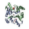 1lqoC S: Starting model for refinement C: citing same article ( |
|---|---|
| Similar structure data |


-Links
 PDBj
PDBj

- Assembly
| Deposited unit | 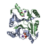
| ||||||||
|---|---|---|---|---|---|---|---|---|---|
| 1 |
| ||||||||
| Unit cell |
| ||||||||
| Details | The biological assembly is a dimer of chains A and B |
-Components
| #1: Protein | Mass: 15143.006 Da / Num. of mol.: 2 Source method: isolated from a genetically manipulated source Source: (gene. exp.) Pseudomonas aeruginosa (bacteria) / Gene: FOSA / Plasmid: PET-20 / Species (production host): Escherichia coli / Production host: #2: Chemical |   Mass: 39.098 Da / Num. of mol.: 2 / Source method: obtained synthetically / Formula: K Mass: 39.098 Da / Num. of mol.: 2 / Source method: obtained synthetically / Formula: K#3: Chemical |   Mass: 54.938 Da / Num. of mol.: 2 / Source method: obtained synthetically / Formula: Mn Mass: 54.938 Da / Num. of mol.: 2 / Source method: obtained synthetically / Formula: Mn#4: Chemical | ChemComp-FCN /   Mass: 138.059 Da / Num. of mol.: 4 / Source method: obtained synthetically / Formula: C3H7O4P / Comment: antibiotic*YM Mass: 138.059 Da / Num. of mol.: 4 / Source method: obtained synthetically / Formula: C3H7O4P / Comment: antibiotic*YM#5: Water | ChemComp-HOH / |  Mass: 18.015 Da / Num. of mol.: 407 / Source method: isolated from a natural source / Formula: H2O Mass: 18.015 Da / Num. of mol.: 407 / Source method: isolated from a natural source / Formula: H2O |
|---|
-Experimental details
-Experiment
| Experiment | Method: X-RAY DIFFRACTION / Number of used crystals: 1 |
|---|
- Sample preparation
| Crystal | Density Matthews: 2.01 Å3/Da / Density % sol: 38.7 % |
|---|---|
| Crystal grow | Temperature: 298 K / Method: vapor diffusion, hanging drop / pH: 7 Details: WELL CONTAINED 40% PENTAERYTHRITOL PROPOXYLATE 629, 0.10M KCL. DROP CONTAINED 0.002 M MNCL2, 0.002M FOSFOMYCIN, 10 MG/ML FOSA, pH 7.0, VAPOR DIFFUSION, HANGING DROP, temperature 298K |
-Data collection
| Diffraction | Mean temperature: 113 K |
|---|---|
| Diffraction source | Source: SYNCHROTRON / Site: APS  / Beamline: 14-BM-C / Wavelength: 0.9 / Beamline: 14-BM-C / Wavelength: 0.9 |
| Detector | Type: ADSC QUANTUM 4 / Detector: CCD / Date: Jun 20, 2002 |
| Radiation | Protocol: SINGLE WAVELENGTH / Monochromatic (M) / Laue (L): M / Scattering type: x-ray |
| Radiation wavelength | Wavelength: 0.9 Å / Relative weight: 1 |
| Reflection | Resolution: 1.19→100 Å / Num. obs: 77910 / % possible obs: 86.3 % / Observed criterion σ(I): -3 / Redundancy: 8.3 % / Rmerge(I) obs: 0.08 / Net I/σ(I): 15 |
| Reflection shell | Resolution: 1.19→1.23 Å / Rmerge(I) obs: 0.31 / Mean I/σ(I) obs: 3.6 / % possible all: 57.8 |
| Reflection | *PLUS Lowest resolution: 20 Å / % possible obs: 90.8 % / Rmerge(I) obs: 0.08 |
| Reflection shell | *PLUS % possible obs: 57.8 % / Rmerge(I) obs: 0.31 |
- Processing
| Software |
| |||||||||||||||||||||||||||||||||
|---|---|---|---|---|---|---|---|---|---|---|---|---|---|---|---|---|---|---|---|---|---|---|---|---|---|---|---|---|---|---|---|---|---|---|
| Refinement | Method to determine structure: MAD Starting model: PDB ENTRY 1LQK Resolution: 1.19→20 Å / Num. parameters: 23565 / Num. restraintsaints: 28753 / Cross valid method: FREE R / σ(F): 0 / Stereochemistry target values: ENGH AND HUBER Details: ANISOTROPIC REFINEMENT REDUCED FREE R (NO CUTOFF) BY 4%
| |||||||||||||||||||||||||||||||||
| Solvent computation | Solvent model: MOEWS & KRETSINGER, J.MOL.BIOL.91(1973)201-228 | |||||||||||||||||||||||||||||||||
| Refine analyze | Num. disordered residues: 6 / Occupancy sum hydrogen: 2007.4 / Occupancy sum non hydrogen: 2576 | |||||||||||||||||||||||||||||||||
| Refinement step | Cycle: LAST / Resolution: 1.19→20 Å
| |||||||||||||||||||||||||||||||||
| Refine LS restraints |
| |||||||||||||||||||||||||||||||||
| Software | *PLUS Name: SHELXL / Version: 97 / Classification: refinement | |||||||||||||||||||||||||||||||||
| Refinement | *PLUS Rfactor obs: 0.147 / Rfactor Rfree: 0.1893 / Rfactor Rwork: 0.1463 | |||||||||||||||||||||||||||||||||
| Solvent computation | *PLUS | |||||||||||||||||||||||||||||||||
| Displacement parameters | *PLUS | |||||||||||||||||||||||||||||||||
| Refine LS restraints | *PLUS
|
