 Movie
Movie Controller


+ Open data

- Basic information
| Entry | Database: PDB / ID: 1qr2 | ||||||
|---|---|---|---|---|---|---|---|
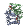
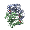
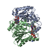
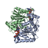
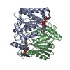
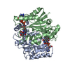
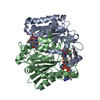
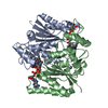
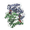
| Title | HUMAN QUINONE REDUCTASE TYPE 2 | ||||||
 Components Components | PROTEIN (QUINONE REDUCTASE TYPE 2) | ||||||
 Keywords Keywords | OXIDOREDUCTASE / QUINONE-REDUCTASE (CYTOSOLIC) / FLAVOPROTEIN / METALLOENZYME | ||||||
| Function / homology |  Function and homology information Function and homology informationribosyldihydronicotinamide dehydrogenase (quinone) / dihydronicotinamide riboside quinone reductase activity / quinone catabolic process / resveratrol binding / oxidoreductase activity, acting on other nitrogenous compounds as donors / melatonin binding / NAD(P)H dehydrogenase (quinone) activity / Phase I - Functionalization of compounds / chloride ion binding / FAD binding ...ribosyldihydronicotinamide dehydrogenase (quinone) / dihydronicotinamide riboside quinone reductase activity / quinone catabolic process / resveratrol binding / oxidoreductase activity, acting on other nitrogenous compounds as donors / melatonin binding / NAD(P)H dehydrogenase (quinone) activity / Phase I - Functionalization of compounds / chloride ion binding / FAD binding / electron transfer activity / oxidoreductase activity / protein homodimerization activity / extracellular exosome / zinc ion binding / nucleoplasm / cytosol Similarity search - Function | ||||||
| Biological species |  Homo sapiens (human) Homo sapiens (human) | ||||||
| Method |  X-RAY DIFFRACTION / MOLECULAR REPLACEMENT / Resolution: 2.1 Å X-RAY DIFFRACTION / MOLECULAR REPLACEMENT / Resolution: 2.1 Å | ||||||
 Authors Authors | Foster, C. / Bianchet, M.A. / Talalay, P. / Amzel, L.M. | ||||||
 Citation Citation | Journal: Biochemistry / Year: 1999 Title: Crystal structure of human quinone reductase type 2, a metalloflavoprotein. Authors: Foster, C.E. / Bianchet, M.A. / Talalay, P. / Zhao, Q. / Amzel, L.M. #1: Journal: Proc.Natl.Acad.Sci.USA / Year: 1997 Title: Unexpected genetic and structural relationships of a long-forgotten flavoenzyme to NAD(P)H:quinone reductase (DT-diaphorase) Authors: Zhao, Q. / Yang, X.L. / Holtzclaw, W.D. / Talalay, P. | ||||||
| History |
|
- Structure visualization
| Structure viewer | Molecule: MolmilJmol/JSmol |
|---|
- Downloads & links
-Download
| PDBx/mmCIF format | 1qr2.cif.gz | 133.5 KB | Display | PDBx/mmCIF format |
|---|---|---|---|---|
| PDB format | pdb1qr2.ent.gz | 103.9 KB | Display | PDB format |
| PDBx/mmJSON format | 1qr2.json.gz | Tree view | PDBx/mmJSON format | |
| Others |  Other downloads Other downloads |
-Validation report
| Arichive directory | https://data.pdbj.org/pub/pdb/validation_reports/qr/1qr2ftp://data.pdbj.org/pub/pdb/validation_reports/qr/1qr2 | HTTPS FTP |
|---|
-Related structure data
| Related structure data |  2qr2C 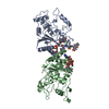 1qrdS S: Starting model for refinement C: citing same article ( |
|---|---|
| Similar structure data |

-Links
 PDBj
PDBj- Assembly
| Deposited unit | 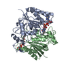
| ||||||||
|---|---|---|---|---|---|---|---|---|---|
| 1 |
| ||||||||
| Unit cell |
| ||||||||
| Noncrystallographic symmetry (NCS) | NCS oper: (Code: given Matrix: (0.761451, -0.007801, -0.648175), Vector: |
-Components
| #1: Protein | Mass: 25849.338 Da / Num. of mol.: 2 Source method: isolated from a genetically manipulated source Source: (gene. exp.) Homo sapiens (human) / Description: SYNTHETIC GENE / Species (production host): Escherichia coli / Cellular location (production host): CYTOPOLASM / Gene (production host): NQO2 / Production host:  #2: Chemical |   Mass: 65.409 Da / Num. of mol.: 2 / Source method: obtained synthetically / Formula: Zn Mass: 65.409 Da / Num. of mol.: 2 / Source method: obtained synthetically / Formula: Zn#3: Chemical |   Mass: 785.550 Da / Num. of mol.: 2 / Source method: obtained synthetically / Formula: C27H33N9O15P2 / Comment: FAD*YM Mass: 785.550 Da / Num. of mol.: 2 / Source method: obtained synthetically / Formula: C27H33N9O15P2 / Comment: FAD*YM#4: Water | ChemComp-HOH / |  Mass: 18.015 Da / Num. of mol.: 194 / Source method: isolated from a natural source / Formula: H2O Mass: 18.015 Da / Num. of mol.: 194 / Source method: isolated from a natural source / Formula: H2OSequence details | THE SEQUENCE DATABASE IS IN ERROR AT RESIDUE 140, CYS. THE CORRECT RESIDUE IS GLY. (RESIDUE 139 IN ...THE SEQUENCE DATABASE IS IN ERROR AT RESIDUE 140, CYS. THE CORRECT RESIDUE IS GLY. (RESIDUE 139 IN THE COORDINATE | |
|---|
-Experimental details
-Experiment
| Experiment | Method: X-RAY DIFFRACTION / Number of used crystals: 1 |
|---|
- Sample preparation
| Crystal | Density Matthews: 2.4 Å3/Da / Density % sol: 49 % | ||||||||||||||||||||||||||||||
|---|---|---|---|---|---|---|---|---|---|---|---|---|---|---|---|---|---|---|---|---|---|---|---|---|---|---|---|---|---|---|---|
| Crystal grow | pH: 7 / Details: pH 7.0 | ||||||||||||||||||||||||||||||
| Crystal | *PLUS | ||||||||||||||||||||||||||||||
| Crystal grow | *PLUS Temperature: 18 ℃ / Method: vapor diffusion, hanging drop | ||||||||||||||||||||||||||||||
| Components of the solutions | *PLUS
|
-Data collection
| Diffraction | Mean temperature: 93 K |
|---|---|
| Diffraction source | Source: ROTATING ANODE / Type: RIGAKU RU200 / Wavelength: 1.5418 |
| Detector | Type: RIGAKU RAXIS IV / Detector: IMAGE PLATE / Date: Jan 1, 1997 |
| Radiation | Monochromator: GRAPHITE / Protocol: SINGLE WAVELENGTH / Monochromatic (M) / Laue (L): M / Scattering type: x-ray |
| Radiation wavelength | Wavelength: 1.5418 Å / Relative weight: 1 |
| Reflection | Resolution: 2.1→100 Å / Num. obs: 27290 / % possible obs: 89.8 % / Redundancy: 6.3 % / Rsym value: 5.7 / Net I/σ(I): 25 |
| Reflection shell | Resolution: 2.1→2.2 Å / Redundancy: 2.6 % / Mean I/σ(I) obs: 4 / Rsym value: 24.4 / % possible all: 75.7 |
| Reflection | *PLUS Num. measured all: 171959 / Rmerge(I) obs: 0.057 |
| Reflection shell | *PLUS % possible obs: 75.7 % / Rmerge(I) obs: 0.244 |
- Processing
| Software |
| ||||||||||||||||||||||||||||||||||||||||||||||||||||||||||||
|---|---|---|---|---|---|---|---|---|---|---|---|---|---|---|---|---|---|---|---|---|---|---|---|---|---|---|---|---|---|---|---|---|---|---|---|---|---|---|---|---|---|---|---|---|---|---|---|---|---|---|---|---|---|---|---|---|---|---|---|---|---|
| Refinement | Method to determine structure: MOLECULAR REPLACEMENT Starting model: PDB ENTRY 1QRD Resolution: 2.1→6 Å / Data cutoff high absF: 1000000 / Data cutoff low absF: 0.001 / Cross valid method: THROUGHOUT / σ(F): 3
| ||||||||||||||||||||||||||||||||||||||||||||||||||||||||||||
| Refine analyze | Luzzati d res low obs: 6 Å | ||||||||||||||||||||||||||||||||||||||||||||||||||||||||||||
| Refinement step | Cycle: LAST / Resolution: 2.1→6 Å
| ||||||||||||||||||||||||||||||||||||||||||||||||||||||||||||
| Refine LS restraints |
| ||||||||||||||||||||||||||||||||||||||||||||||||||||||||||||
| Refine LS restraints NCS | NCS model details: RESTRAINTS | ||||||||||||||||||||||||||||||||||||||||||||||||||||||||||||
| LS refinement shell | Resolution: 2.1→2.15 Å / Total num. of bins used: 15
| ||||||||||||||||||||||||||||||||||||||||||||||||||||||||||||
| Xplor file |
| ||||||||||||||||||||||||||||||||||||||||||||||||||||||||||||
| Software | *PLUS Name: X-PLOR / Classification: refinement | ||||||||||||||||||||||||||||||||||||||||||||||||||||||||||||
| Refinement | *PLUS Lowest resolution: 6 Å / σ(F): 3 / % reflection Rfree: 10 % / Rfactor obs: 0.219 | ||||||||||||||||||||||||||||||||||||||||||||||||||||||||||||
| Solvent computation | *PLUS | ||||||||||||||||||||||||||||||||||||||||||||||||||||||||||||
| Displacement parameters | *PLUS | ||||||||||||||||||||||||||||||||||||||||||||||||||||||||||||
| Refine LS restraints | *PLUS
| ||||||||||||||||||||||||||||||||||||||||||||||||||||||||||||
| LS refinement shell | *PLUS Rfactor Rfree: 0.329 / % reflection Rfree: 10 % / Rfactor Rwork: 0.299 |
