 Movie
Movie Controller


+ Open data

- Basic information
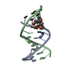
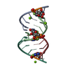
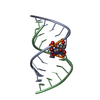
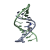
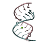
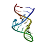
| Entry | Database: PDB / ID: 1qph | ||||||||||||||||||
|---|---|---|---|---|---|---|---|---|---|---|---|---|---|---|---|---|---|---|---|
| Title | CRYSTAL STRUCTURE OF THE A-DNA DODECAMER GACCACGTGGTC | ||||||||||||||||||
 Components Components | 5'-D(P* Keywords KeywordsDNA / DOUBLE HELIX / A-DNA / MAX PROTEIN / DNA CURVATURE | Function / homology | DNA / DNA (> 10) |  Function and homology information Function and homology informationMethod |  X-RAY DIFFRACTION / RIGID BODY REFINEMENT / Resolution: 2.5 Å X-RAY DIFFRACTION / RIGID BODY REFINEMENT / Resolution: 2.5 Å  Authors AuthorsRaaijmakers, H. / Suck, D. / Mayer, C. |  CitationJournal: To be Published CitationJournal: To be PublishedTitle: Crystal Structure Analysis of the A-DNA Dodecamer GACCACGTGGTC Authors: Raaijmakers, H. / Suck, D. / Mayer, C. History |
|
- Structure visualization
| Structure viewer | Molecule: MolmilJmol/JSmol |
|---|
- Downloads & links
-Download
| PDBx/mmCIF format | 1qph.cif.gz | 16.8 KB | Display | PDBx/mmCIF format |
|---|---|---|---|---|
| PDB format | pdb1qph.ent.gz | 10.2 KB | Display | PDB format |
| PDBx/mmJSON format | 1qph.json.gz | Tree view | PDBx/mmJSON format | |
| Others |  Other downloads Other downloads |
-Validation report
| Arichive directory | https://data.pdbj.org/pub/pdb/validation_reports/qp/1qphftp://data.pdbj.org/pub/pdb/validation_reports/qp/1qph | HTTPS FTP |
|---|
-Related structure data
| Similar structure data | |
|---|---|
| Other databases |
|

-Links
 PDBj
PDBj


- Assembly
| Deposited unit | 
| ||||||||||
|---|---|---|---|---|---|---|---|---|---|---|---|
| 1 | 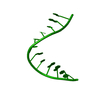
| ||||||||||
| Unit cell |
| ||||||||||
| Components on special symmetry positions |
|
-Components
| #1: DNA chain | Mass: 3663.392 Da / Num. of mol.: 1 / Source method: obtained synthetically / Details: RECOGNITION SEQUENCE OF MAX PROTEIN |
|---|---|
| #2: Water | ChemComp-HOH /  Mass: 18.015 Da / Num. of mol.: 45 / Source method: isolated from a natural source / Formula: H2O Mass: 18.015 Da / Num. of mol.: 45 / Source method: isolated from a natural source / Formula: H2O |
-Experimental details
-Experiment
| Experiment | Method: X-RAY DIFFRACTION / Number of used crystals: 1 |
|---|
- Sample preparation
| Crystal | Density Matthews: 3.07 Å3/Da / Density % sol: 59.88 % | ||||||||||||
|---|---|---|---|---|---|---|---|---|---|---|---|---|---|
| Crystal grow | Temperature: 277 K / Method: vapor diffusion, hanging drop / pH: 6.5 Details: (NH4)2SO4, WATER, pH 6.5, VAPOR DIFFUSION, HANGING DROP, temperature 277.0K | ||||||||||||
| Components of the solutions |
|
-Data collection
| Diffraction | Mean temperature: 278 K |
|---|---|
| Diffraction source | Source: ROTATING ANODE / Type: MACSCIENCE / Wavelength: 1.5418 |
| Detector | Type: MARRESEARCH / Detector: IMAGE PLATE / Date: Sep 25, 1996 |
| Radiation | Protocol: SINGLE WAVELENGTH / Monochromatic (M) / Laue (L): M / Scattering type: x-ray |
| Radiation wavelength | Wavelength: 1.5418 Å / Relative weight: 1 |
| Reflection | Resolution: 2.5→23 Å / Num. all: 1781 / Num. obs: 1781 / % possible obs: 97.7 % / Observed criterion σ(I): 0 / Redundancy: 3.6 % / Biso Wilson estimate: 19.7 Å2 / Rmerge(I) obs: 0.054 / Net I/σ(I): 43.1 |
- Processing
| Software |
| ||||||||||||||||||||||||||||||||||||||||||||||||||||||||||||
|---|---|---|---|---|---|---|---|---|---|---|---|---|---|---|---|---|---|---|---|---|---|---|---|---|---|---|---|---|---|---|---|---|---|---|---|---|---|---|---|---|---|---|---|---|---|---|---|---|---|---|---|---|---|---|---|---|---|---|---|---|---|
| Refinement | Method to determine structure: RIGID BODY REFINEMENT Starting model: NDB ID ADL045 Resolution: 2.5→7 Å / σ(F): 2 Stereochemistry target values: G. PARKINSON, J. VOJTECHOVSKY, L. CLOWNEY, A.T. BRUNGER, H.M. BERMAN, NEW PARAMETERS FOR THE REFINEMENT OF NUCLEIC ACID CONTAINING STRUCTURES, ACTA CRYST. D, 52, 57-64 (1996). Details: SIMULATED ANNEALING REFINEMENT
| ||||||||||||||||||||||||||||||||||||||||||||||||||||||||||||
| Refinement step | Cycle: LAST / Resolution: 2.5→7 Å
| ||||||||||||||||||||||||||||||||||||||||||||||||||||||||||||
| Refine LS restraints |
| ||||||||||||||||||||||||||||||||||||||||||||||||||||||||||||
| LS refinement shell | Resolution: 2.5→2.61 Å / Total num. of bins used: 8
| ||||||||||||||||||||||||||||||||||||||||||||||||||||||||||||
| Xplor file | Serial no: 1 / Param file: PARAM_NDBX3.DNA / Topol file: TOP_NDBX3.DNA |
