 Movie
Movie Controller

[English] 日本語
 Yorodumi
- PDB-1qmi: Crystal structure of RNA 3'-terminal phosphate cyclase, an ubiqui... -
+ Open data

- Basic information
| Entry | Database: PDB / ID: 1qmi | ||||||
|---|---|---|---|---|---|---|---|
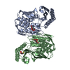
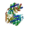
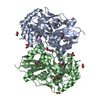
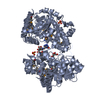
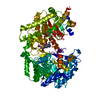
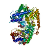
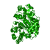
| Title | Crystal structure of RNA 3'-terminal phosphate cyclase, an ubiquitous enzyme with unusual topology | ||||||
 Components Components | RNA 3'-TERMINAL PHOSPHATE CYCLASE | ||||||
 Keywords Keywords | LIGASE / 2'3'CYCLIC PHOSPHATE RNA | ||||||
| Function / homology |  Function and homology information Function and homology informationRNA 3'-terminal-phosphate cyclase (ATP) / RNA-3'-phosphate cyclase activity / RNA processing / ATP binding / cytoplasm Similarity search - Function | ||||||
| Biological species |  | ||||||
| Method |  X-RAY DIFFRACTION / SYNCHROTRON / MAD / Resolution: 2.8 Å X-RAY DIFFRACTION / SYNCHROTRON / MAD / Resolution: 2.8 Å | ||||||
 Authors Authors | Palm, G.J. / Billy, E. / Filipowicz, W. / Wlodawer, A. | ||||||
 Citation Citation | Journal: Structure / Year: 2000 Title: Crystal Structure of RNA 3'-Terminal Phosphate Cyclase, a Ubiquitous Enzyme with Unusual Topology Authors: Palm, G.J. / Billy, E. / Filipowicz, W. / Wlodawer, A. | ||||||
| History |
|
- Structure visualization
| Structure viewer | Molecule: MolmilJmol/JSmol |
|---|
- Downloads & links
-Download
| PDBx/mmCIF format | 1qmi.cif.gz | 228.1 KB | Display | PDBx/mmCIF format |
|---|---|---|---|---|
| PDB format | pdb1qmi.ent.gz | 190.3 KB | Display | PDB format |
| PDBx/mmJSON format | 1qmi.json.gz | Tree view | PDBx/mmJSON format | |
| Others |  Other downloads Other downloads |
-Validation report
| Arichive directory | https://data.pdbj.org/pub/pdb/validation_reports/qm/1qmiftp://data.pdbj.org/pub/pdb/validation_reports/qm/1qmi | HTTPS FTP |
|---|
-Related structure data


-Links
 PDBj
PDBj
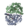
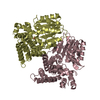
- Assembly
| Deposited unit | 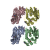
| ||||||||||||||||
|---|---|---|---|---|---|---|---|---|---|---|---|---|---|---|---|---|---|
| 1 | 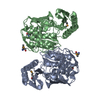
| ||||||||||||||||
| 2 | 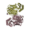
| ||||||||||||||||
| Unit cell |
| ||||||||||||||||
| Noncrystallographic symmetry (NCS) | NCS oper:
|
-Components
| #1: Protein | Mass: 37248.703 Da / Num. of mol.: 4 Source method: isolated from a genetically manipulated source Source: (gene. exp.) References: UniProt: P46849, RNA 3'-terminal-phosphate cyclase (ATP) Has protein modification | Y | Sequence details | C-TERMINAL HIS TAG ADDED | |
|---|
-Experimental details
-Experiment
| Experiment | Method: X-RAY DIFFRACTION / Number of used crystals: 1 |
|---|
- Sample preparation
| Crystal | Density Matthews: 2.74 Å3/Da / Density % sol: 55 % | ||||||||||||||||||||||||||||||
|---|---|---|---|---|---|---|---|---|---|---|---|---|---|---|---|---|---|---|---|---|---|---|---|---|---|---|---|---|---|---|---|
| Crystal grow | pH: 5.5 Details: THE PROTEIN WAS CRYSTALLIZED FROM 13-15% MPEG2000, 200 MM NA-CITRATE PH 4.0, 200 MM TRIS/HCL PH 8.0, 2 MM DTT. PROTEIN CONCENTRATION WAS CA. 15 MG/ML. | ||||||||||||||||||||||||||||||
| Crystal grow | *PLUS Temperature: 20 ℃ / Method: vapor diffusion, hanging drop | ||||||||||||||||||||||||||||||
| Components of the solutions | *PLUS
|
-Data collection
| Diffraction | Mean temperature: 105 K |
|---|---|
| Diffraction source | Source: SYNCHROTRON / Site: NSLS  / Beamline: X9B / Wavelength: 0.97942 / Beamline: X9B / Wavelength: 0.97942 |
| Detector | Type: MARRESEARCH / Detector: IMAGE PLATE / Date: Jan 28, 1998 |
| Radiation | Protocol: SINGLE WAVELENGTH / Monochromatic (M) / Laue (L): M / Scattering type: x-ray |
| Radiation wavelength | Wavelength: 0.97942 Å / Relative weight: 1 |
| Reflection | Resolution: 2.8→20 Å / Num. obs: 41596 / % possible obs: 96.9 % / Observed criterion σ(I): -3 / Redundancy: 3.9 % / Rmerge(I) obs: 0.045 / Net I/σ(I): 17.8 |
| Reflection shell | Resolution: 2.8→2.9 Å / Redundancy: 3.8 % / Rmerge(I) obs: 0.198 / Mean I/σ(I) obs: 7.5 / % possible all: 91.6 |
| Reflection | *PLUS Num. measured all: 154068 |
| Reflection shell | *PLUS % possible obs: 91.6 % |
- Processing
| Software |
| ||||||||||||||||||||||||||||||||||||||||||||||||||||||||||||
|---|---|---|---|---|---|---|---|---|---|---|---|---|---|---|---|---|---|---|---|---|---|---|---|---|---|---|---|---|---|---|---|---|---|---|---|---|---|---|---|---|---|---|---|---|---|---|---|---|---|---|---|---|---|---|---|---|---|---|---|---|---|
| Refinement | Method to determine structure: MAD / Resolution: 2.8→10 Å / Rfactor Rfree error: 0.008 / Data cutoff high absF: 100000 / Data cutoff low absF: 0.01 / Cross valid method: THROUGHOUT / σ(F): 2 Details: MODEL WAS NOT FULLY REFINED, THE HIS TAG IN CHAINS A,B,C,D WERE NOT VISIBLE IN THE ELECTRON DENSITY. THE CIS PEPTIDE GLY 232 - PRO 233 COULD ONLY BE MODELLED IN THE FULLY REFINED STRUCTURE 1QMH
| ||||||||||||||||||||||||||||||||||||||||||||||||||||||||||||
| Displacement parameters | Biso mean: 38.3 Å2 | ||||||||||||||||||||||||||||||||||||||||||||||||||||||||||||
| Refinement step | Cycle: LAST / Resolution: 2.8→10 Å
| ||||||||||||||||||||||||||||||||||||||||||||||||||||||||||||
| Refine LS restraints |
| ||||||||||||||||||||||||||||||||||||||||||||||||||||||||||||
| Refine LS restraints NCS | NCS model details: RESTRAINTS ONLY ON COORDINATES / Rms dev position: 0.48 Å / Weight position: 200 | ||||||||||||||||||||||||||||||||||||||||||||||||||||||||||||
| LS refinement shell | Resolution: 2.8→2.92 Å / Rfactor Rfree error: 0.034 / Total num. of bins used: 8
|
